Tobias Spronk1,2,3, Oliver Kraff1, Gregor Schaefers3,4, and Harald H Quick1,2
1Erwin L. Hahn Institute for MR Imaging, University of Duisburg-Essen, Essen, Germany, 2High-Field and Hybrid MR Imaging, University Hospital Essen, Essen, Germany, 3MRI-STaR Magnetic Resonance Institute for Safety, Technology and Research GmbH, Gelsenkirchen, Germany, 4MR:comp GmbH, Testing Services for MR Safety & Compatibility, Gelsenkirchen, Germany
1Erwin L. Hahn Institute for MR Imaging, University of Duisburg-Essen, Essen, Germany, 2High-Field and Hybrid MR Imaging, University Hospital Essen, Essen, Germany, 3MRI-STaR Magnetic Resonance Institute for Safety, Technology and Research GmbH, Gelsenkirchen, Germany, 4MR:comp GmbH, Testing Services for MR Safety & Compatibility, Gelsenkirchen, Germany
This study presents a
numerical simulation approach that predicts the MR image artifact size of
passive medical implants with a high accuracy.
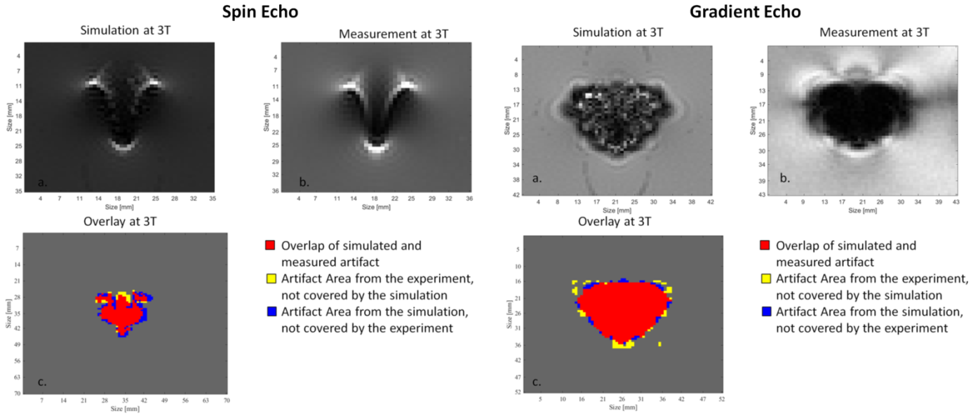
(A) Simulated spin echo and gradient echo images of a
titanium rod acquired on a 3 T MRI
system. (B) Measured spin echo and
gradient echo image of the titanium rod. (C) Overlay
of the artifacts from image (A) and
(B). Red color: both artifacts are congruent; yellow: measured artifact
only; blue: simulated artifact only.
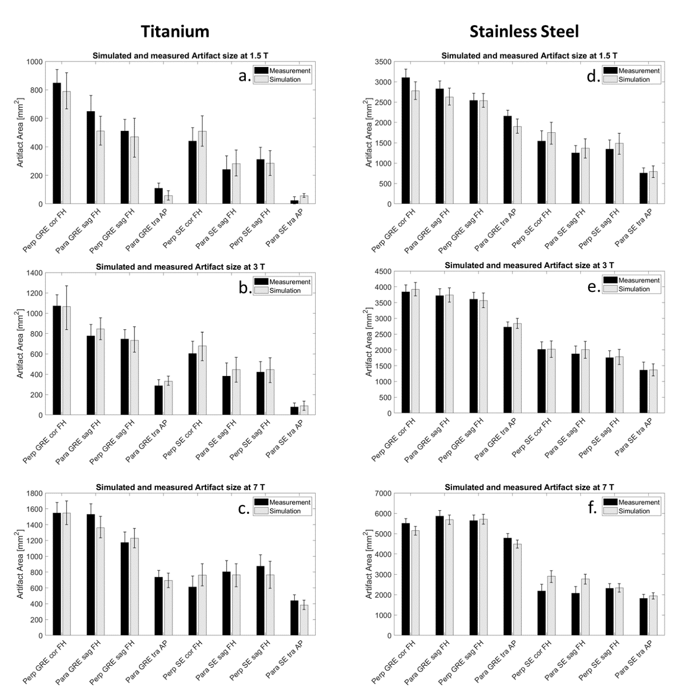
This figure compares the artifact size of the titanium
(A-C) and stainless steel (D-F) rod of the simulations (light grey) and the
measurements (black) in mm2 separated by the different field
strengths, (A,D) 1.5 T, (B,E) 3 T, (C,F) 7 T. Artifact sizes are shown for
different implant orientations in regard to B0 (para: parallel and
perp: perpendicular), sequences (GRE and SE), slice orientations in regard to
implant orientation (cor, sag, tra), and phase-encoding direction (FH, AP).