Kübra Keskin1, Brian Hargreaves2,3,4, and Krishna S. Nayak1,5
1Electrical and Computer Engineering, University of Southern California, Los Angeles, CA, United States, 2Radiology, Stanford University, Stanford, CA, United States, 3Electrical Engineering, Stanford University, Stanford, CA, United States, 4Bioengineering, Stanford University, Stanford, CA, United States, 5Biomedical Engineering, University of Southern California, Los Angeles, CA, United States
1Electrical and Computer Engineering, University of Southern California, Los Angeles, CA, United States, 2Radiology, Stanford University, Stanford, CA, United States, 3Electrical Engineering, Stanford University, Stanford, CA, United States, 4Bioengineering, Stanford University, Stanford, CA, United States, 5Biomedical Engineering, University of Southern California, Los Angeles, CA, United States
We demonstrate a pipeline for realistic simulation of
in-vivo MRI around metal. We use this
simulator to predict the performance of multi-spectral imaging on emerging
low-field systems (e.g. 0.55 Tesla) and predict optimal imaging parameters.
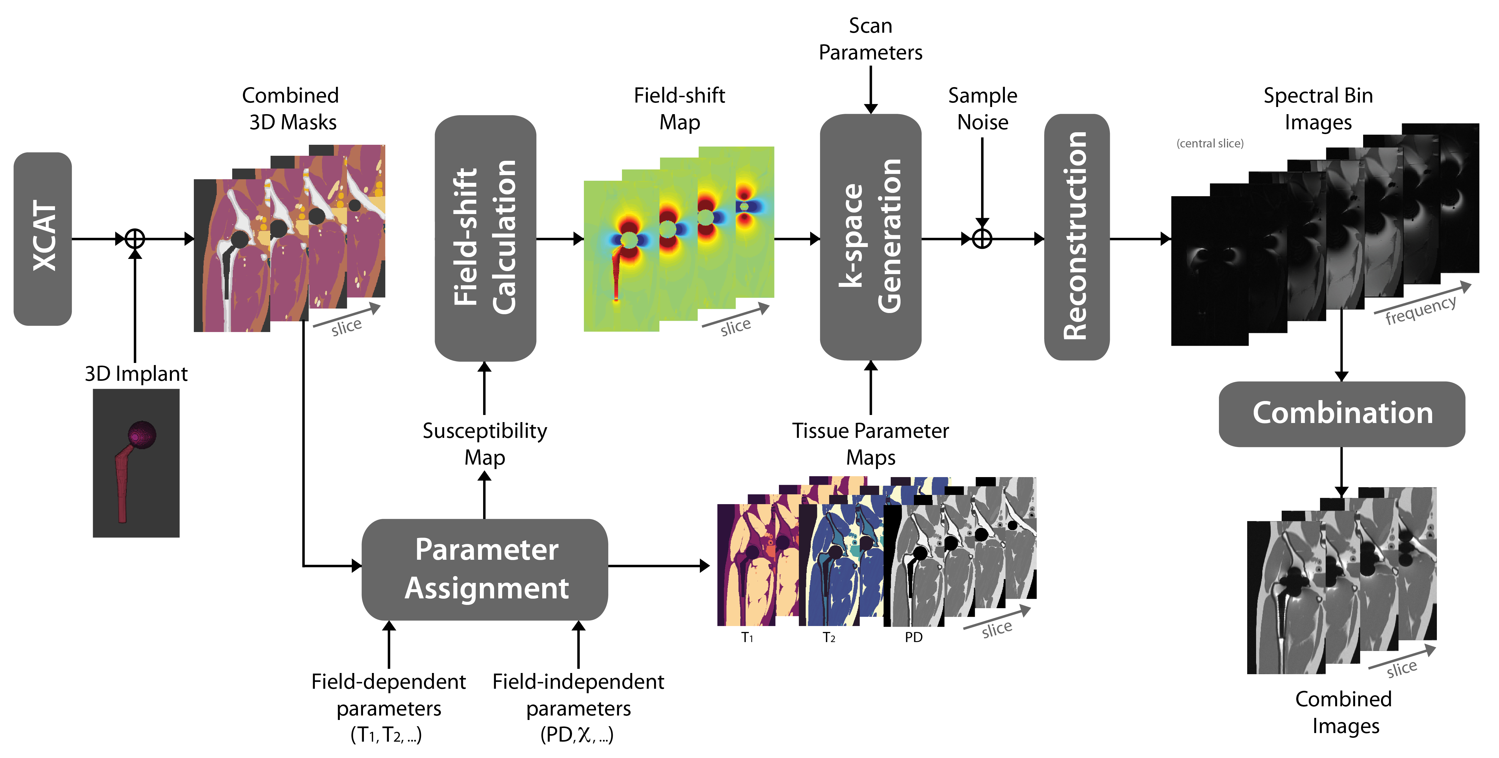
Figure 1: Simulation
Pipeline. 3D XCAT body and implant masks are utilized to create tissue
parameter maps. The susceptibility map is used to calculate the field shift. K-space
is simulated using the field shift, proton density (PD) and MSI sequence
parameters with phase encoding in both left-right and anterior-posterior
directions. Complex Gaussian noise is added in k-space. Spectral bin images are
reconstructed and combined with quadrature summation.
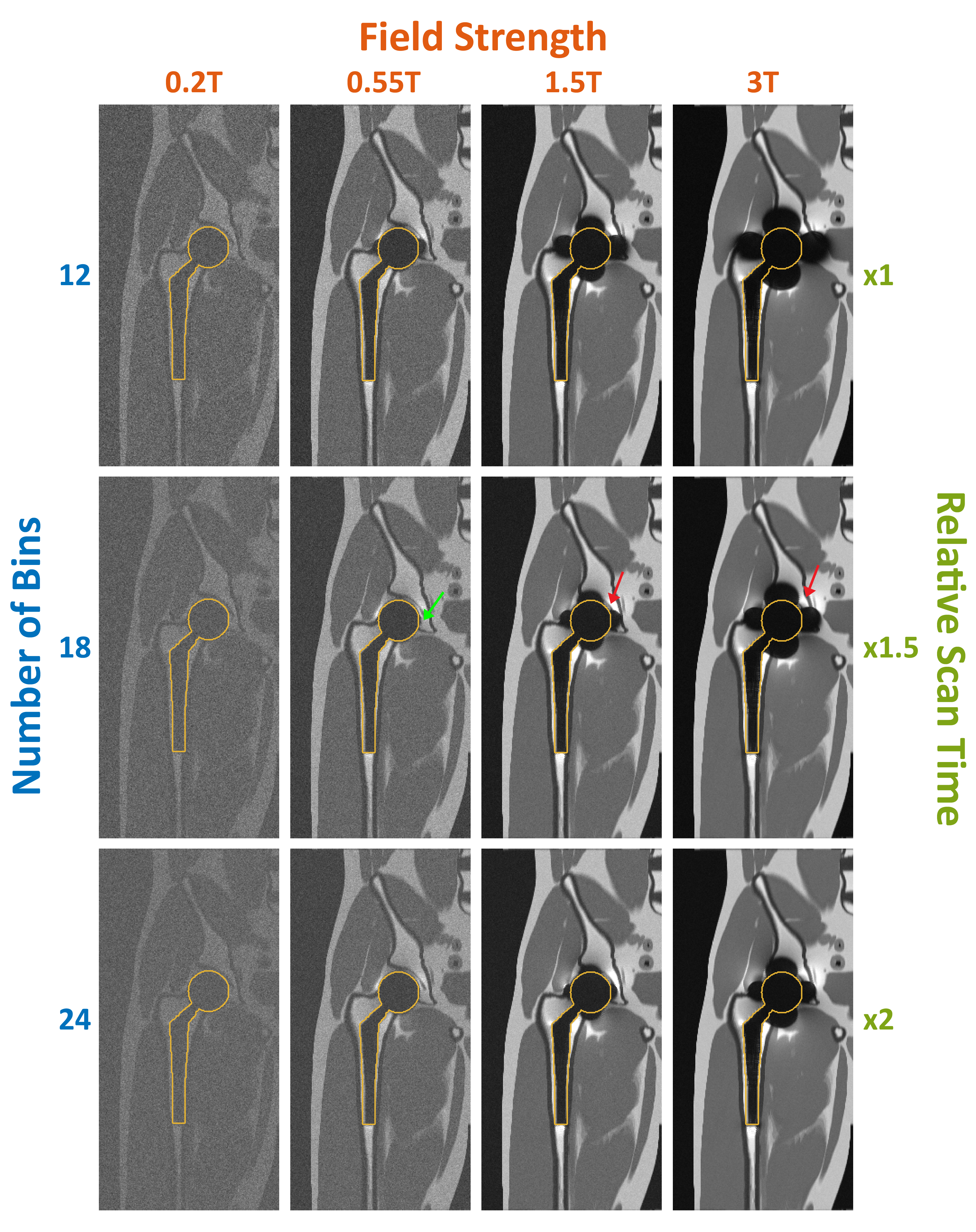
Figure 2: MAVRIC-SL simulation of total hip arthroplasty. Central coronal slices from 3D volumes are shown for B0 = 0.2T,
0.55T, 1.5T, 3T, and Nbins = 12, 18, 24. Other parameters: BW/pixel = 625Hz;
RF bandwidth = 2.25kHz and bin spacing 1kHz. Implant contours are shown with yellow
outlines. Artifacts around the implant are indicated with red arrows. A small artifact
adjacent to implant surface at 0.55T is shown with green arrow. Total scan times
are 3:53, 5:49, and 7:45 for Nbins of 12, 18, and 24, respectively.