Alexander R Toews1,2, Daehyun Yoon1, and Brian A Hargreaves1,2,3
1Radiology, Stanford University, Stanford, CA, United States, 2Electrical Engineering, Stanford University, Stanford, CA, United States, 3Bioengineering, Stanford University, Stanford, CA, United States
1Radiology, Stanford University, Stanford, CA, United States, 2Electrical Engineering, Stanford University, Stanford, CA, United States, 3Bioengineering, Stanford University, Stanford, CA, United States
Conventional multispectral imaging mitigates geometric distortion near metal at the expense of readout blur and residual intensity artifacts. Phantom and simulation results demonstrate the proposed method reduces readout blur and in some cases eliminates residual intensity artifacts.
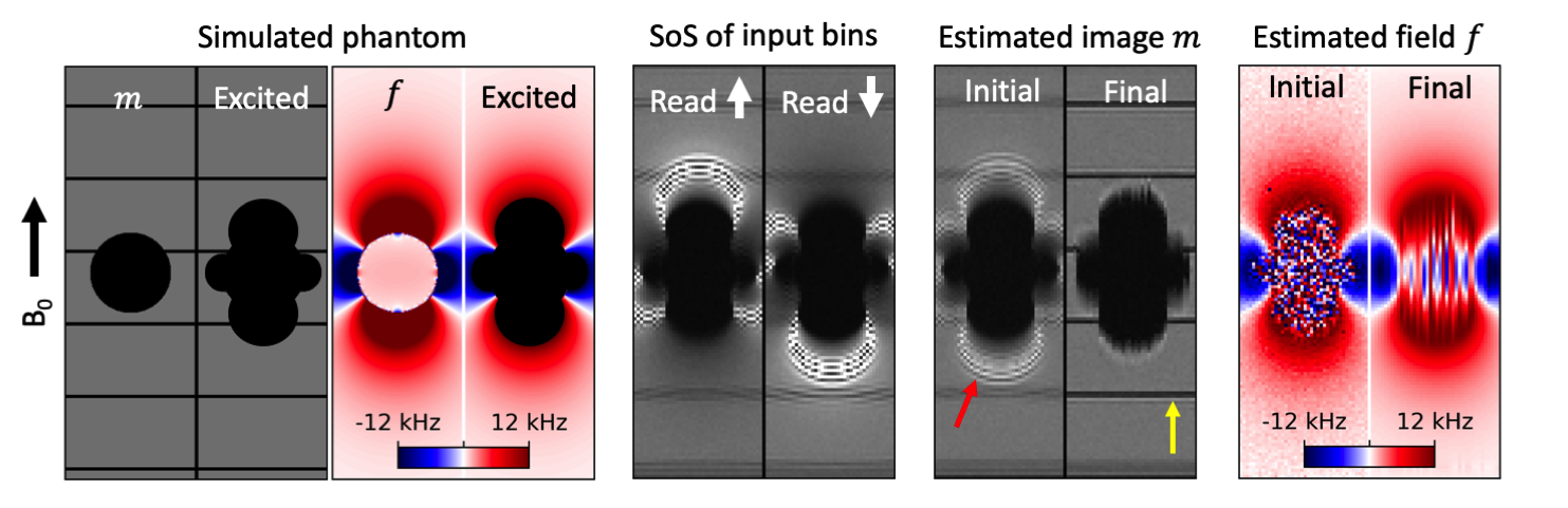
Figure 3: Simulated phantom study of a ball with magnetic susceptibility 900 ppm at 3T. Input images exhibit readout blur and ripple artifact due to VAT. Ripple artifacts are reduced in the initial image (red arrow) and eliminated in the final estimation of $$$m$$$. Grid lines are sharper in the final image (yellow arrow), indicating mitigation of the VAT blur effect. Final $$$f$$$ agrees well with the true field within the region of excited spins.
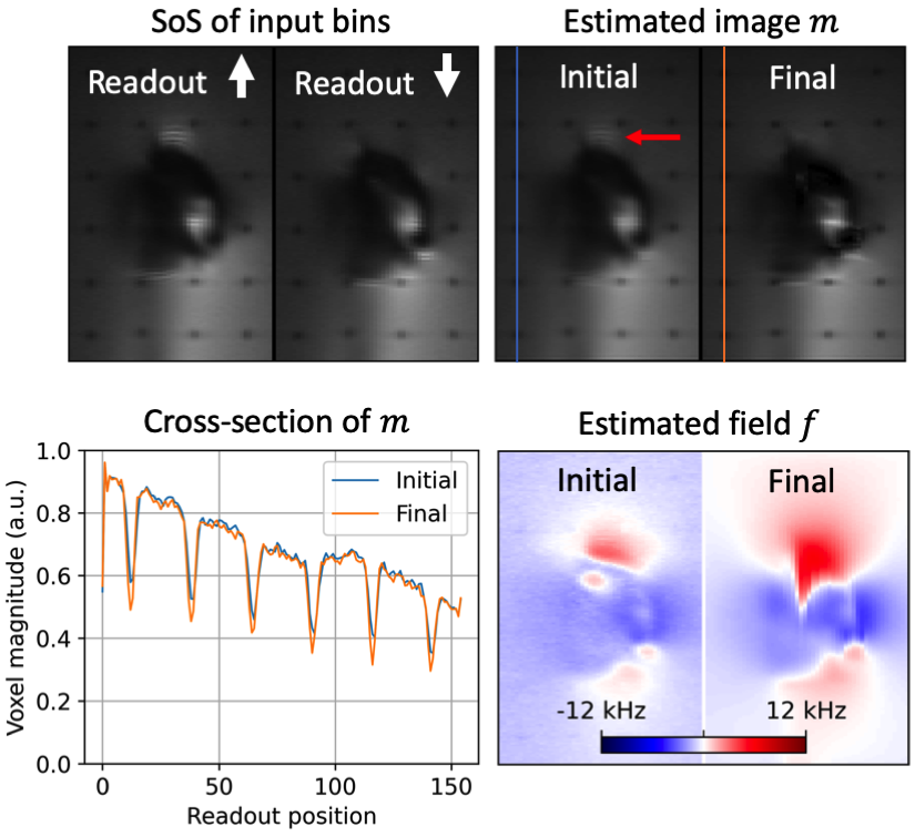
Figure 5: Results in shoulder phantom shown for an off-center coronal slice. Ripple-like intensity artifacts are apparent in the input and initial images (red arrow), but significantly reduced in the final image. Differences in readout resolution are less apparent than in Figure 4 due to the absence of grid lines.