Kain Kyle1,2 and Chenyu Wang1,2
1Sydney Neuroimaging Analysis Centre, Sydney, Australia, 2University of Sydney, Sydney, Australia
1Sydney Neuroimaging Analysis Centre, Sydney, Australia, 2University of Sydney, Sydney, Australia
In this investigation we compared bias correction techniques by estimating brain volume change in healthy controls. We found are proposed iterative WM N4 bias correction technique resulted in the lowest percentage brain volume change.
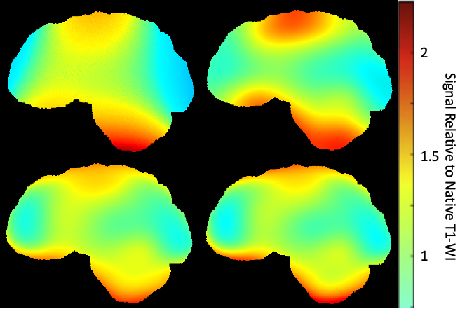
Figure 1. Example of bias correction applied to native T1-WI. Maps were generated by dividing the corrected image by the native image. Bias correction methods N3 (top left), N4 (top right), N4W(bottom left) and N4ITER(bottom right).
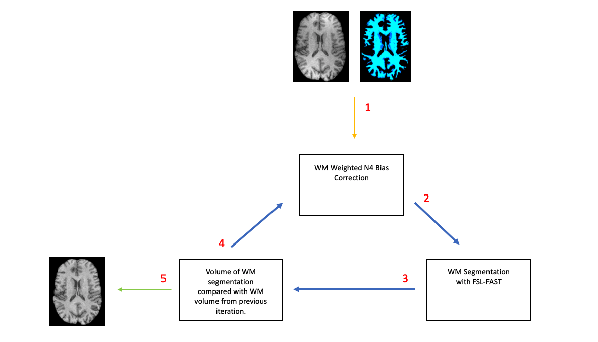
Figure 2. Summary of iterative WM N4 bias correction technique. 1) Initial WM mask is generated with FSL-FAST on native T1-WI and used for N4Wcorrection. 2) WM mask is segmented from N4 corrected image with FSL-FAST. 3) Volume of WM mask from step 2, is compared to the volume of the WM mask from the previous iteration, or initial input WM mask if it is the first iteration. 4) If the change in volume is greater than 0.05%, the updated mask is used to perform N4Won the native T1-WI. 5) If the change in volume is less than 0.05 percent, the process is terminated.