Junlan Lu1, Suphachart Leewiwatwong2, David Mummy3, Elianna Bier2, and Bastiaan Driehuys3
1Medical Physics, Duke University, Durham, NC, United States, 2Biomedical Engineering, Duke University, Durham, NC, United States, 3Radiology, Duke University, Durham, NC, United States
1Medical Physics, Duke University, Durham, NC, United States, 2Biomedical Engineering, Duke University, Durham, NC, United States, 3Radiology, Duke University, Durham, NC, United States
The
short imaging time of hyperpolarized 129Xe MRI imposes a constraint
to image resolution. This can be alleviated using CNNs to enhance low-resolution
ventilation imaging features. Quantitative SNR and SSIM analysis indicate
significant improvement in SNR and structural similarity.
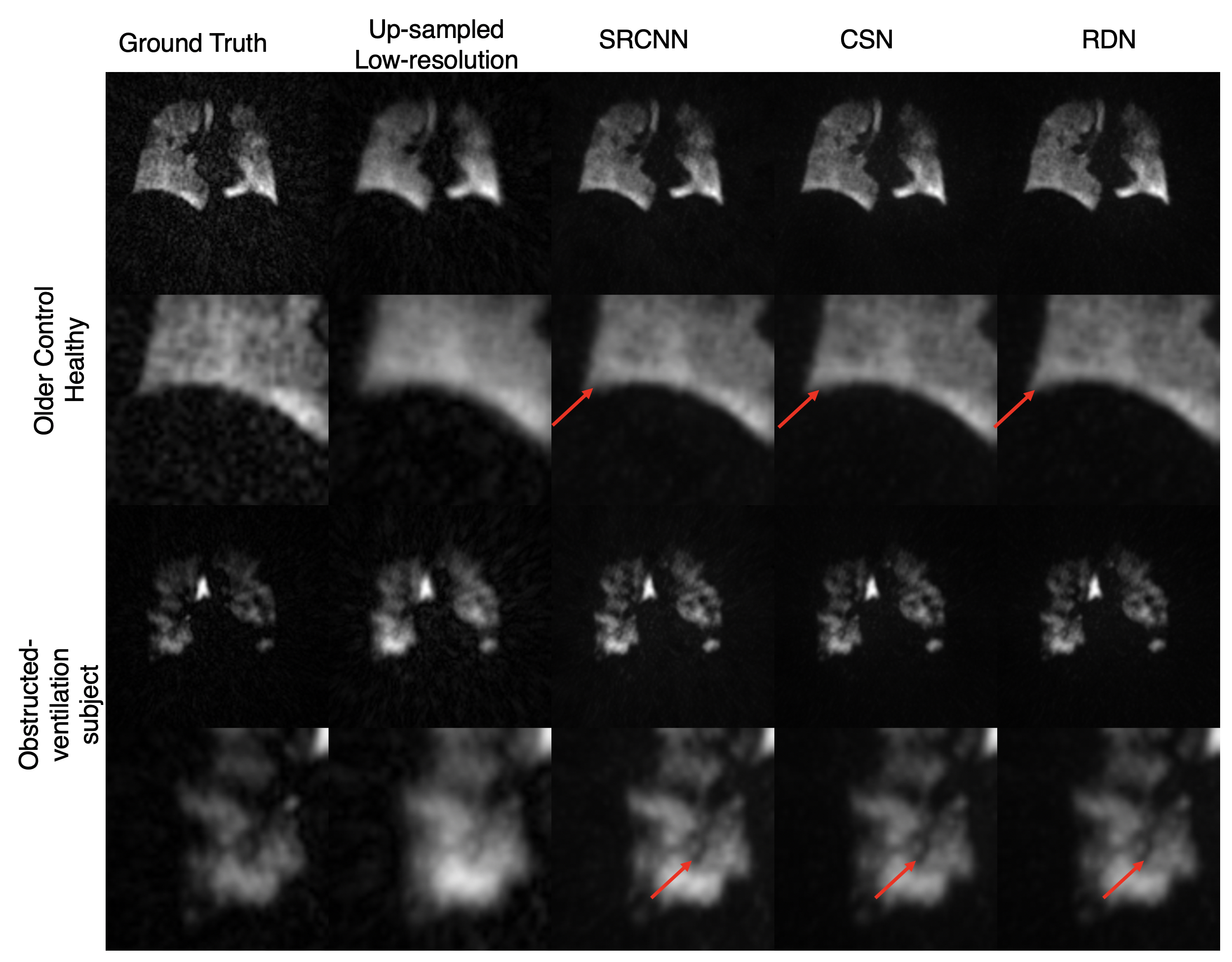
Fig. 3) The
visual effect of the various models trained on the k-space removal size 128x128
dataset on a healthy subject (rows 1-2) and one with visible ventilation
defects (rows 3-4). Regions of interests are highlighted to show differences in
image texture, image noise, and feature sharpness. Row 2 indicates that edges
are sharpened while decreasing background noise. Row 4 indicates the recovery
of the ventilation defect.
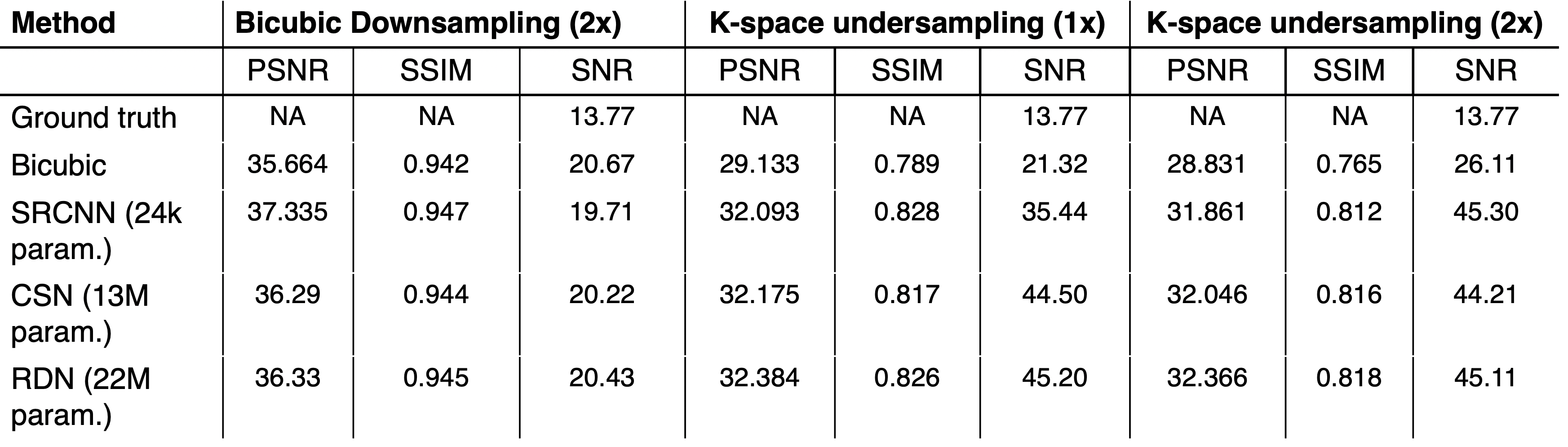
Fig. 4) Benchmark
results (PSNR/SSIM/SNR) of all experiments. For models trained with datasets generated
from k-space under-sampling (columns 2,3), improvements are seen in all three
metrics compared to the bicubic up-sampling method on the low-resolution image.
Moreover, SNR of all model outputs are higher than that of the original ground
truth. However, for models trained with datasets generated from bicubic down-sampling
of the high-resolution image (column 1), SNR is decreased because the models
are able to accurately reproduce the noise pattern.