Joshua R Astley1,2, Alberto M Biancardi1, Paul J Hughes1, Laurie J Smith1, Helen Marshall1, Guilhem J Collier1, James Eaden1, Nicholas D Weatherley1, Jim M Wild1, and Bilal A Tahir1,2
1POLARIS, University of Sheffield, Sheffield, United Kingdom, 2Oncology and Metabolism, University of Sheffield, Sheffield, United Kingdom
1POLARIS, University of Sheffield, Sheffield, United Kingdom, 2Oncology and Metabolism, University of Sheffield, Sheffield, United Kingdom
We compared the performance of several 3D convolutional neural network architectures and loss functions for segmentation of ventilated lungs on a large and diverse multi-nuclear hyperpolarized gas MRI dataset.
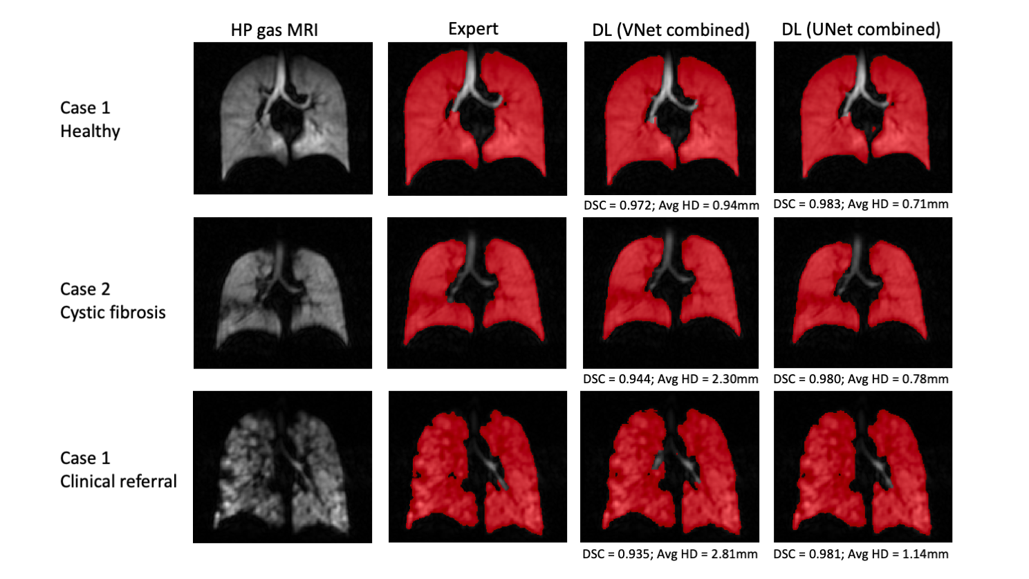
Figure 3. Example coronal slices of the UNet and VNet combined 3He and 129Xe trained segmentations for three cases with different diseases compared to the expert segmentations. DSC and Avg HD values are given for each case.
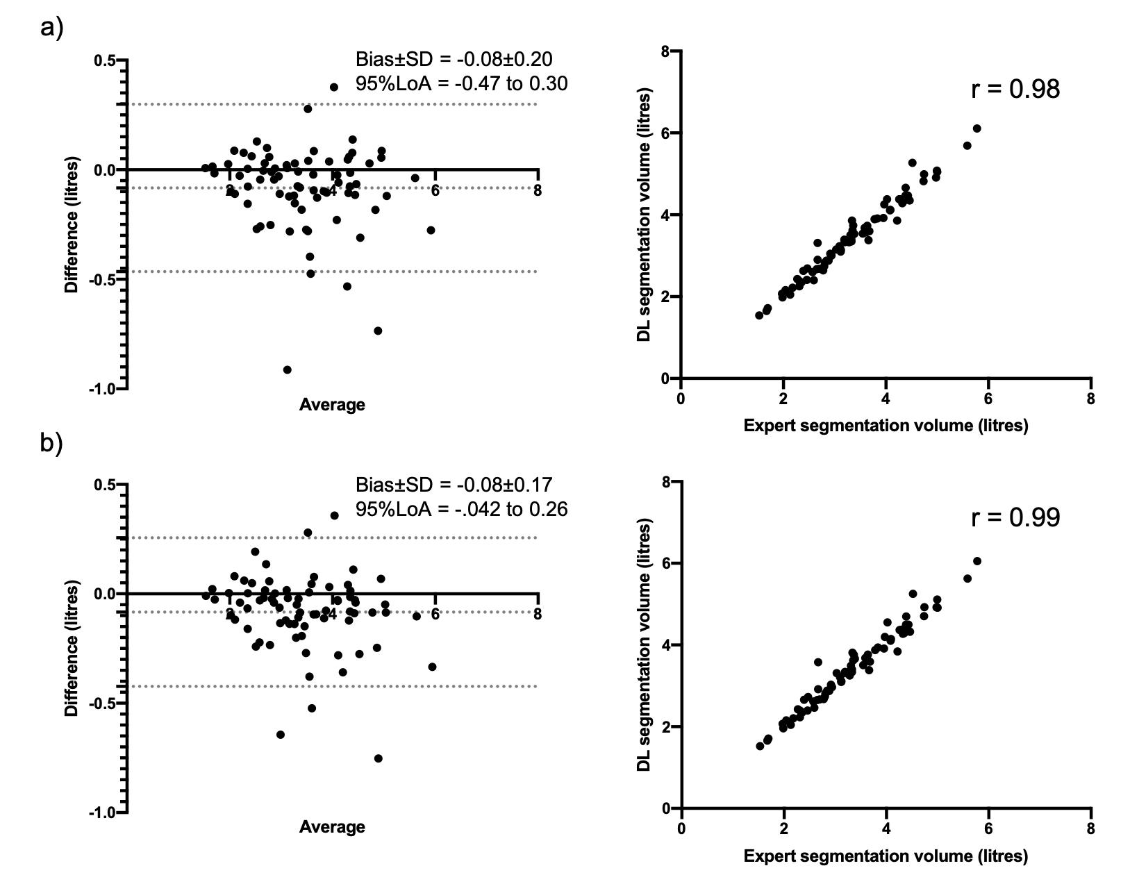
Figure 4. Correlation and agreement analysis of lung volumes for 75 testing set cases compared to expert segmentations for combined 3He and 129Xe a) VNet and b) UNet models.