Rolf F Schulte1, Guilhem J Collier2, James Ball2, Graham Norquay2, Madhwesha Rao2, and Jim M Wild2
1GE Healthcare, Munich, Germany, 2POLARIS, Department of Infection Immunity & Cardiovascular Disease, University of Sheffield, Sheffield, United Kingdom
1GE Healthcare, Munich, Germany, 2POLARIS, Department of Infection Immunity & Cardiovascular Disease, University of Sheffield, Sheffield, United Kingdom
- Density-weighted MRSI: SNR and encoding efficient way to detect xenon gas in alveoli, lung tissue and blood
- A frequency-tailored RF excitation pulse excites the gas phase with 0.1° flip and tissue/blood signal with 10° reliably
- Resulting quantitative ratio maps are of good SNR and high quality
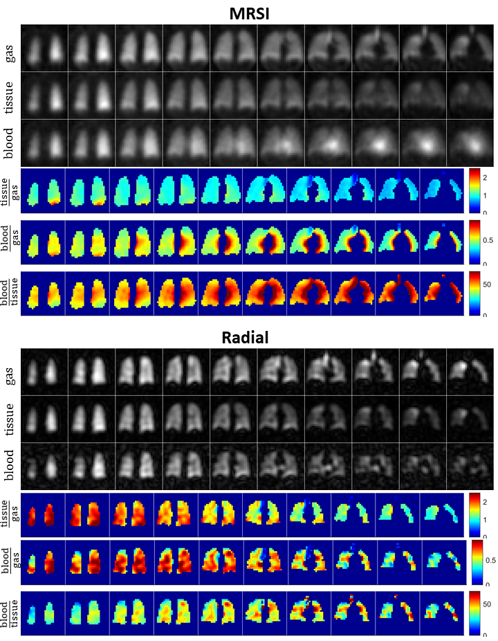
Fig. 4: Volunteer 1: Gas, tissue, blood and its corresponding ratio maps.
The heart is visible in the MRSI, but not radial blood maps. Ratio maps in
percent are scaled to 2.5%, 0.9% and 70% for tissue/gas, blood/gas and
blood/tissue ratios, respectively.
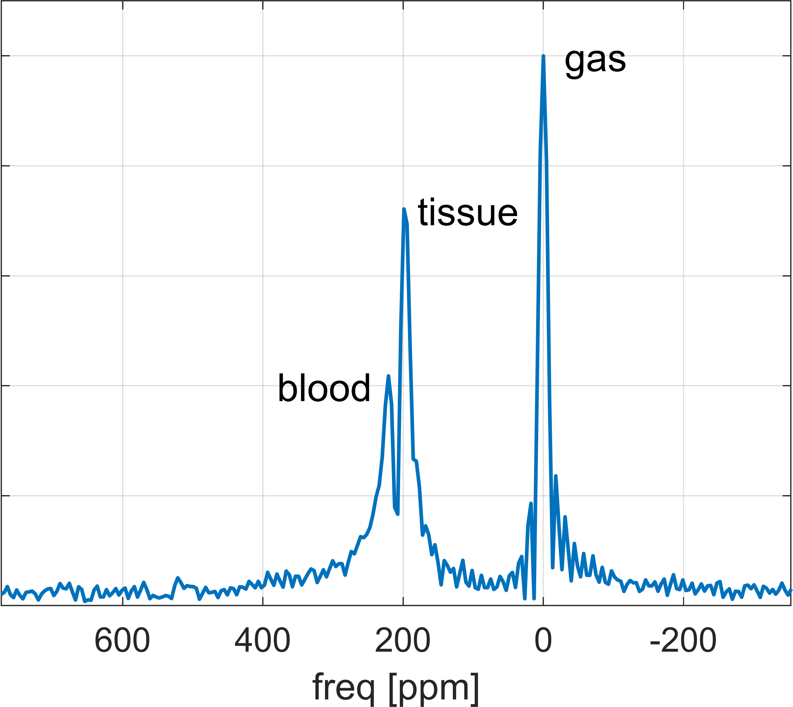
Fig. 3: Representative spectrum from volunteer
1 (x=13, y=16, z=7). Besides spectral zero-filling to 256 points and spatial
zero-filling by a factor of 2, no further post-processing steps (such as
apodisation or denoising) were performed. Some Gibb’s ringing is visible around
the gas peak due to the longer T2*. This exemplifies the good spectral quality
and excellent SNR.