Madhwesha R Rao1, Guilhem J Collier1, Graham Norquay1, Rolf F Schulte2, and Jim M Wild1
1University of Sheffield, Sheffield, United Kingdom, 2GE Healthcare, Munich, Germany
1University of Sheffield, Sheffield, United Kingdom, 2GE Healthcare, Munich, Germany
1. 3D density-weighted MR spectroscopic images of the human brain with hyperpolarized 129Xe produced images with slice thickness of 2 cm and interpolated slice thickness of 0.625 cm.
2. The achieved slice thickness is more than a factor of 2 thinner than those reported earlier.
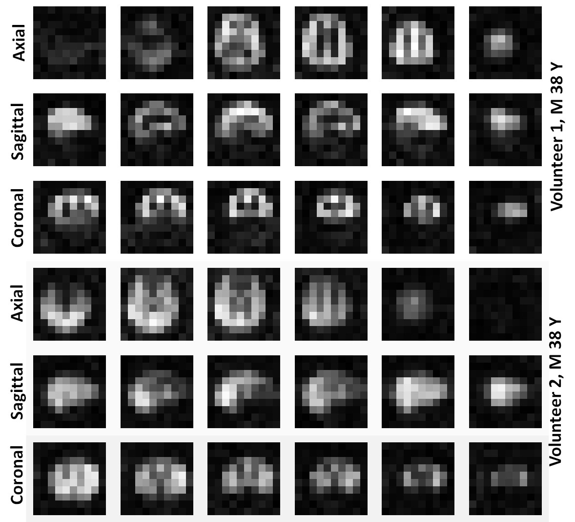
Figure 3: Images of HP 129Xe xenon dissolved in the grey matter for both volunteers at the acquired slice thickness of 2 cm.
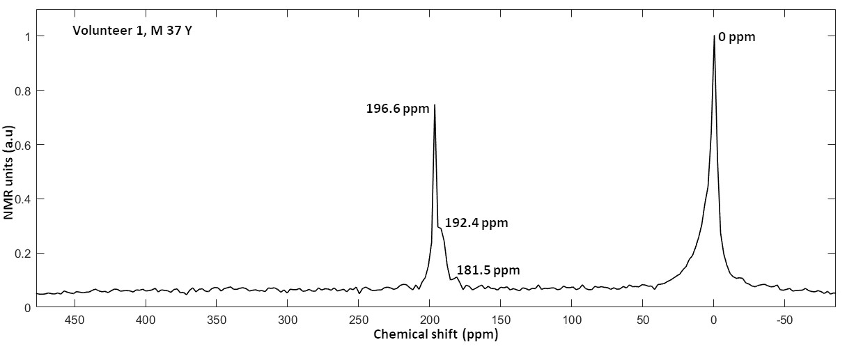
Figure 2: Average spectrum of all the spatially resolved 1101 RF-acquisitions from Volunteer 1 (Male 37 years).