Kai Ruppert1, Faraz Amzajerdian1, Yi Xin1, Hooman Hamedani1, Luis Loza1, Tahmina S Achekzai1, Ryan J Baron1, Ian F Duncan1, Harrilla Profka1, Yiwen Qian1, Stephen Kadlecek1, Alessandra Fusco2, Benjamin Sinder3, Patrick J Cahill3, Brian Snyder3,4, Thomas P Schaer2, and Rahim R Rizi1
1University of Pennsylvania, Philadelphia, PA, United States, 2School of Veterinary Medicine, University of Pennsylvania, Kennett Square, PA, United States, 3Children's Hospital of Philadelphia, Philadelphia, PA, United States, 4Boston Children's Hospital, Boston, MA, United States
1University of Pennsylvania, Philadelphia, PA, United States, 2School of Veterinary Medicine, University of Pennsylvania, Kennett Square, PA, United States, 3Children's Hospital of Philadelphia, Philadelphia, PA, United States, 4Boston Children's Hospital, Boston, MA, United States
In a rib-tether rabbit model, dynamic 1D
simultaneous dissolved- and gas-phase hyperpolarized xenon-129 MRI measurements were found
to be sensitive to pulmonary abnormalities secondary to thoracic insufficiency
syndrome.
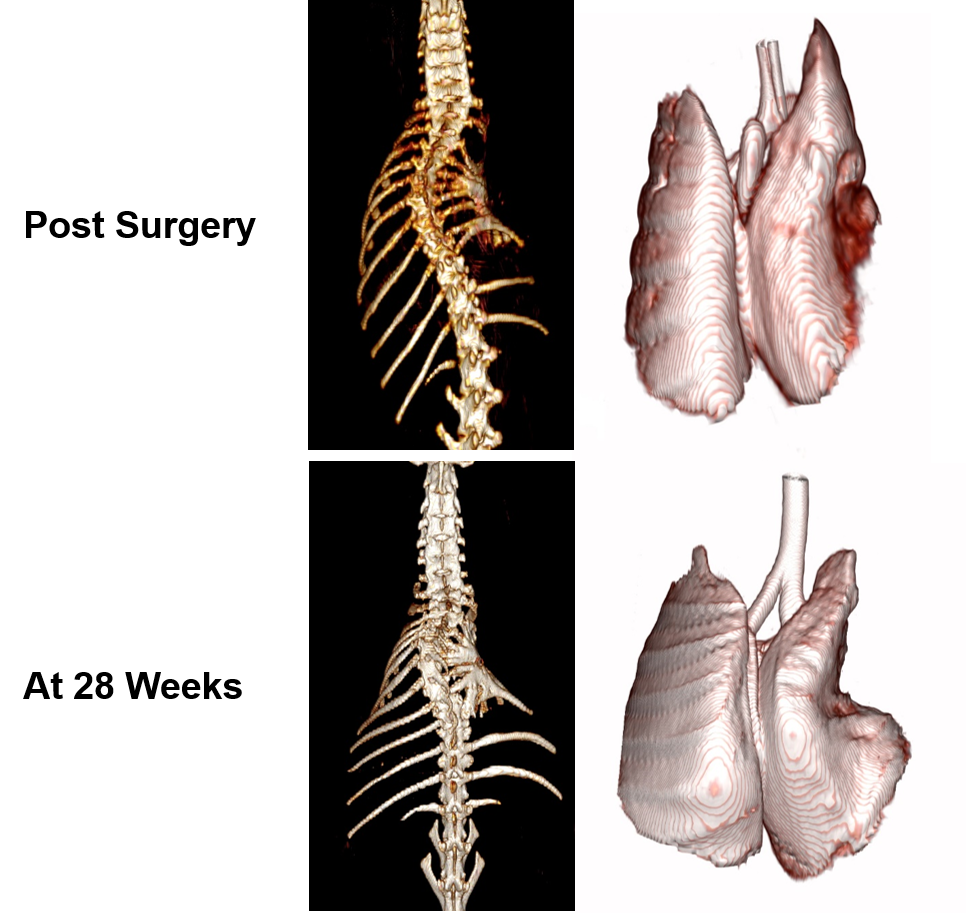
Figure 1. CT renderings of the
spine (left column) and the lungs (right column) in a rib-tether rabbit model immediately
after surgery (6 weeks of age, top row) and at
22 weeks post surgery (28 weeks of age, bottom row) of age. The
successful model implementation resulted in a spinal deformation that highly restricted
the expansion of the right lung during maturation.
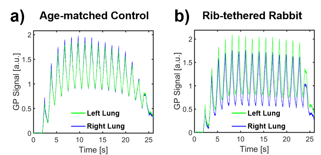
Figure 2. GP signal dynamics
during multi-breath 1D projection acquisitions aggregated for the left and
right lung. (a) In the age-matched control rabbit, both lungs are ventilated
symmetrically throughout the respiratory cycle. (b) In the rib-tethered rabbit, the
restricted right lung is more poorly ventilated than the left lung, most likely
due to decreased compliance.