Yurii Shepelytskyi1,2, Vira Grynko1,2, Tao Li3, Ayman Hassan4,5, Karl Granberg4, and Mitchell S Albert2,3,5
1Chemistry and Materials Science Program, Lakehead University, Thunder Bay, ON, Canada, 2Thunder Bay Regional Health Research Institute, Thunder Bay, ON, Canada, 3Chemistry, Lakehead University, Thunder Bay, ON, Canada, 4Thunder Bay Regional Health Sciences Centre, Thunder Bay, ON, Canada, 5Northern Ontario School of Medicine, Thunder Bay, ON, Canada
1Chemistry and Materials Science Program, Lakehead University, Thunder Bay, ON, Canada, 2Thunder Bay Regional Health Research Institute, Thunder Bay, ON, Canada, 3Chemistry, Lakehead University, Thunder Bay, ON, Canada, 4Thunder Bay Regional Health Sciences Centre, Thunder Bay, ON, Canada, 5Northern Ontario School of Medicine, Thunder Bay, ON, Canada
A substantial
reduction in the signal variability of HP 129Xe dissolved in the
brain due to the application of the initial 90O depolarization
radiofrequency pulse was demonstrated. This concept can be implemented in all HP 129Xe
dissolved phase imaging.
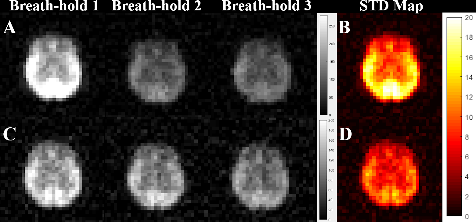
Figure
2. (A) HP 129Xe brain images of
representative healthy volunteer acquired using a GRE imaging 9 s into the
breath-hold without an initial depolarization pulse. (B) Standard SNR deviation
map calculated based on images (A). (C) HP GRE 129Xe brain images
acquired 9 s after the initial depolarization pulse. (D) Standard SNR deviation
map calculated for (C) images. It can be clearly seen that the application of the
initial depolarization pulse yields lower signal variability
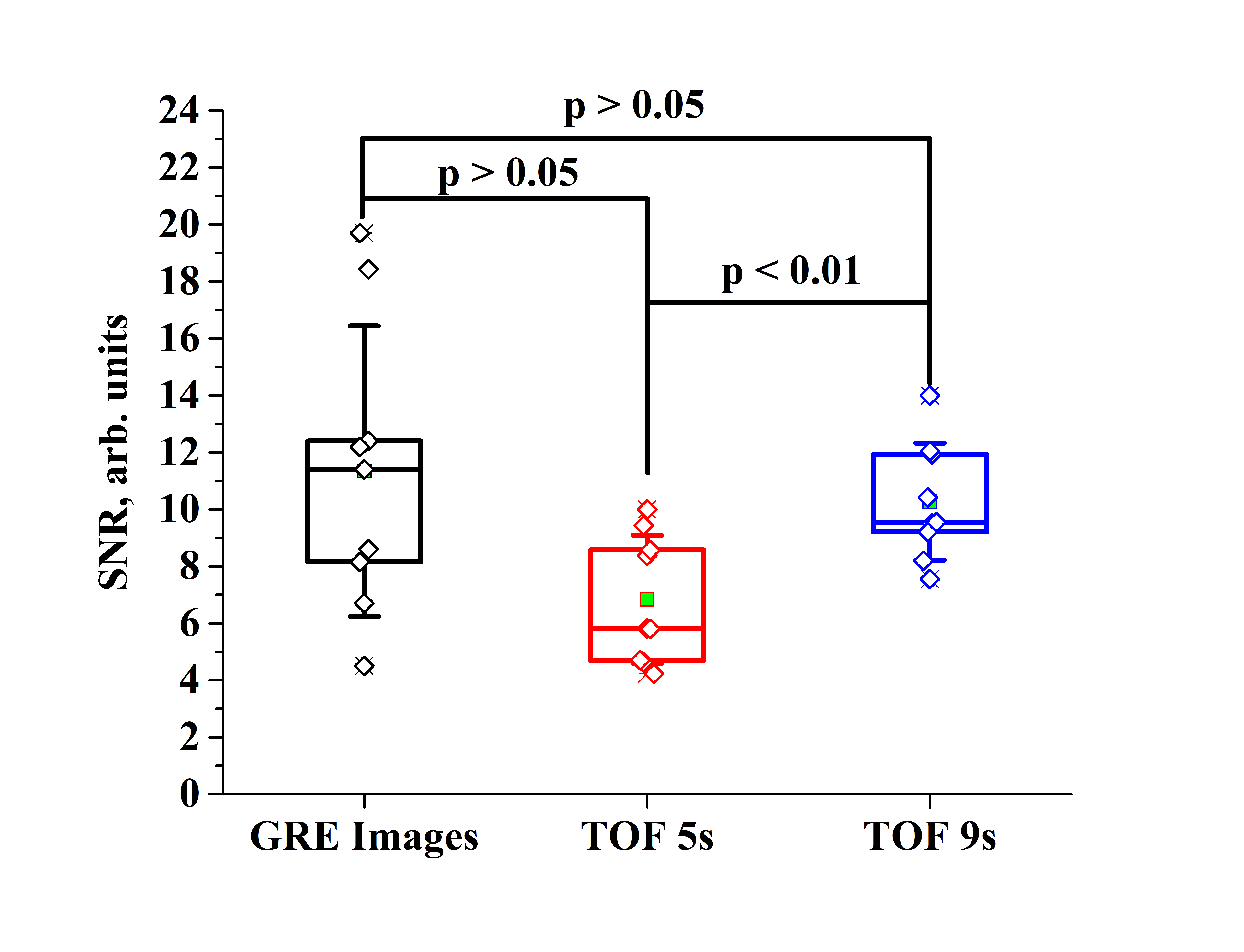
Figure
4. Comparative box-chart of the image SNR. The black
box represents the SNR of the GRE images acquired 9s into the breath-hold
without an initial depolarization radiofrequency pulse. The red box represents the
SNR of the GRE images acquired 5s after the initial depolarization pulse. The
blue box corresponds to the SNR of the GRE images acquired 9s after the initial
depolarization pulse.