Shingo Matsumoto1, Neil J. Stewart1, Hitomi Nakano1, Takuya Hashimoto2, and Hiroshi Hirata1
1Information Science and Technologies, Hokkaido University, Sapporo, Japan, 2Department of Chemistry, Chiba University, Chiba, Japan
1Information Science and Technologies, Hokkaido University, Sapporo, Japan, 2Department of Chemistry, Chiba University, Chiba, Japan
We demonstrated the feasibility of in vivo cell death imaging by 13C MRI of hyperpolarized [1-13C]fumarate, prepared by parahydrogen-induced polarization (PHIP) a low-cost alternative of typical dDNP, in acetaminophen-induced hepatitis model mouse.
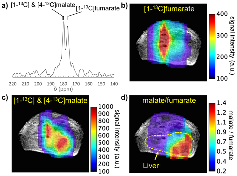
Figure 4. In
vivo
CSI of hyperpolarized [1-13C]fumarate
metabolism in an acetaminophen-induced hepatitis mouse. (a) Representative 13C NMR
spectrum of hyperpolarized [1-13C]fumarate
and its metabolite at the liver. (b) map of hyperpolarized [1-13C]fumarate
CSI signal intensity overlaid on an
anatomical 1H MRI
image. (c) map of hyperpolarized [1-13C]
& [4-13C]malate.
(d) parametric map of the malate/fumarate ratio; a biomarker of cellular
necrosis.
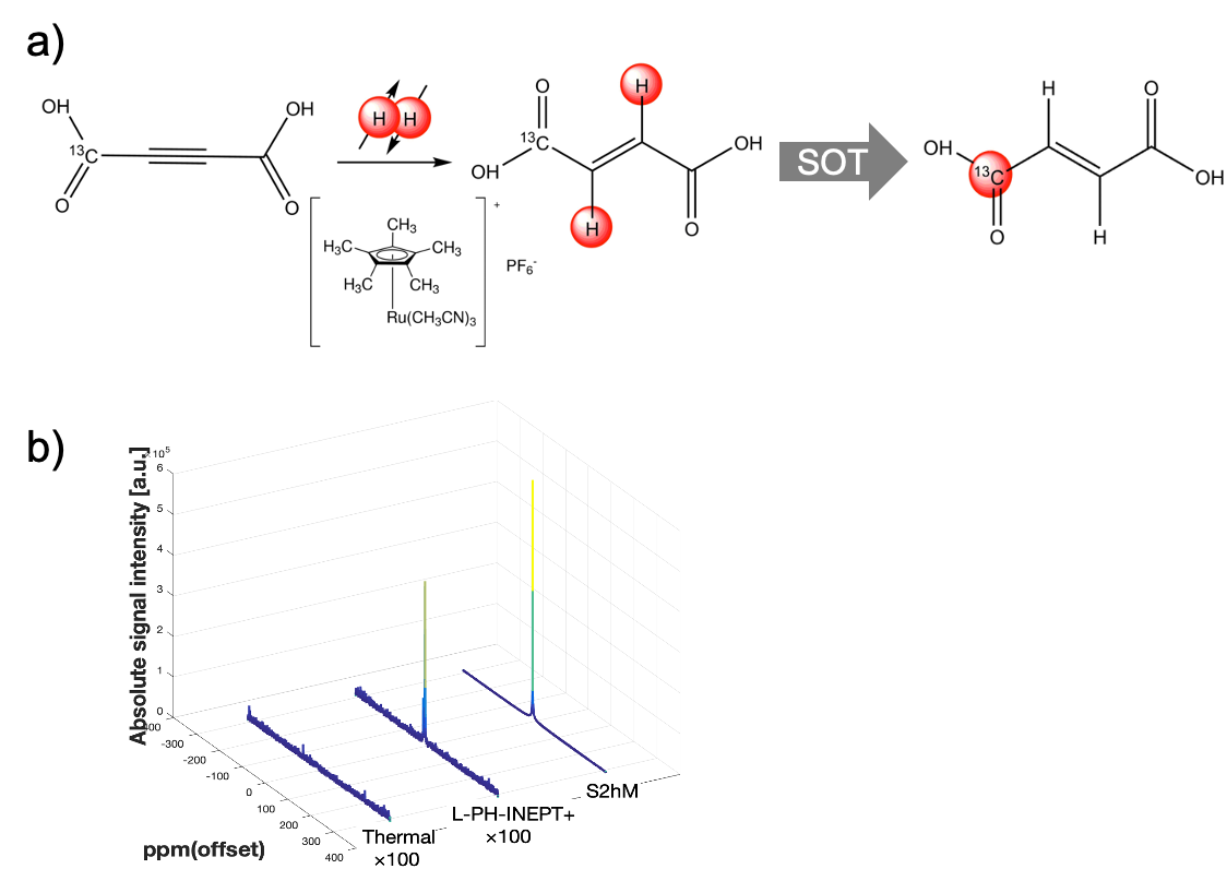
Figure 2. (a)
Preparation
of hyperpolarized [1-13C]fumarate
by trans-alkenylation
with parahydrogen.
(b) 13C
NMR of hyperpolarized [1-13C]
fumarate using different 1H-to-13C spin order transfer pulse sequences.