Jasmine Y Graham1, Adam W Autry2, Yaewon Kim2, Robert A Bok2, Yan Li2, Peder EZ Larson1,2, Daniel B Vigneron1,2, and Jeremy W Gordon2
1Bioengineering, UC San Francisco, UC Berkeley, San Francisco, CA, United States, 2Radiology and Biomedical Imaging, UC San Francisco, San Francisco, CA, United States
1Bioengineering, UC San Francisco, UC Berkeley, San Francisco, CA, United States, 2Radiology and Biomedical Imaging, UC San Francisco, San Francisco, CA, United States
Higher resolution 7.5 mm2 HP [1-13C]pyruvate EPI images improved kinetic rate quantification in human brain by reducing partial volume effects. Selected voxels near blood vessels and in white and gray matter showed >20% higher kPL with higher resolution pyruvate images.
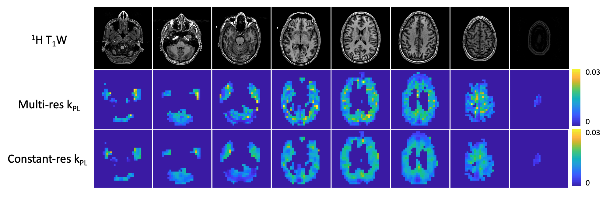
Figure 2. Kinetic rate maps for pyruvate to lactate conversion, using 15 × 15 mm2 lactate signals with 7.5 × 7.5 mm2 pyruvate and synthetic 15 × 15 mm2 pyruvate images thresholded to SNR >5 and <30% fit error, along with reference proton images. The constant-resolution kPL map shows smoothing of the kinetic rates in the brain as compared to the multi-resolution kPL map.
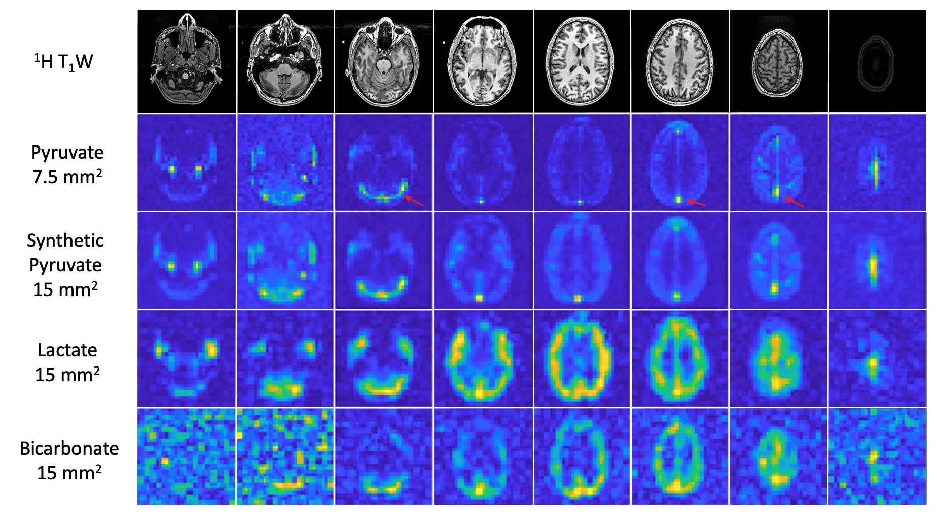
Figure 1. Hyperpolarized 13C pyruvate, lactate and bicarbonate signals summed over 60 seconds with reference proton images. The synthetic 15 × 15 mm2 pyruvate data set was obtained by convolving the 7.5 × 7.5 mm2 pyruvate data with an averaging kernel. The red arrows point out the high pyruvate signal in the transverse sinus for the third slice and in the superior sagittal sinus for the sixth and seventh slices.