Hyeong Yeon Lee1, Najmiddin Mamadjonov2, Nguyen Trong Nguyen2, Luu-Ngoc Do3, Tien Ahn Nguyen3, and Ilwoo Park3,4,5
1Emergency Medicine, Chonnam National University Hospital, Gwangju, Korea, Republic of, 2Biomedical Science, Chonnam National University, Gwangju, Korea, Republic of, 3Radiology, Chonnam National University, Gwangju, Korea, Republic of, 4Radiology, Chonnam National University Hospital, Gwangju, Korea, Republic of, 5Artificial Intelligence Convergence, Chonnam National University, Gwangju, Korea, Republic of
1Emergency Medicine, Chonnam National University Hospital, Gwangju, Korea, Republic of, 2Biomedical Science, Chonnam National University, Gwangju, Korea, Republic of, 3Radiology, Chonnam National University, Gwangju, Korea, Republic of, 4Radiology, Chonnam National University Hospital, Gwangju, Korea, Republic of, 5Artificial Intelligence Convergence, Chonnam National University, Gwangju, Korea, Republic of
This study demonstrated that hyperpolarized 13C metabolic imaging can identify the metabolic changes within 1 hour of return of spontaneous circulation in rodent model of ventricular fibrillation-induced cardiac arrest.
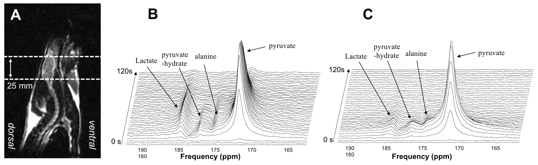
Figure 1. Examples of time-resolved 13C dynamic spectra from a 25 mm slice of rat brain from a CA model (B) and a sham control (C). Hyperpolarized 13C MR spectroscopy data revealed that the cardiac arrest model produced a high level of pyruvate-to-lactate conversion while the sham control had a smaller level of pyruvate-to-lactate conversion compared to the CA model.
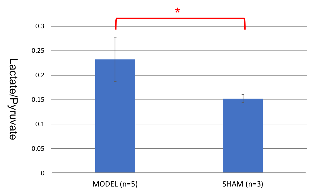
Figure 2. The ratio of lactate to pyruvate in the CA model was statistically higher than that in the sham control (P=0.025).