Yurie Shirai1, Michinobu Nagao1, Noriko Kikuchi1, Atsushi Yamamoto1, Yuka Matsuo1, Risako Nakao1, Kiyoe Ando1, Eri Watanabe1, Shinichi Nunoda1, Masami Yoneyama2, and Syuji Sakai1
1Tokyo Women’s Medical University, Tokyo, Japan, 2Philips Japan, Tokyo, Japan
1Tokyo Women’s Medical University, Tokyo, Japan, 2Philips Japan, Tokyo, Japan
Native T1 mapping can be minimally invasive and have high diagnostic accuracy for CAV after heart transplantation, with no contrast and radiation exposure required.
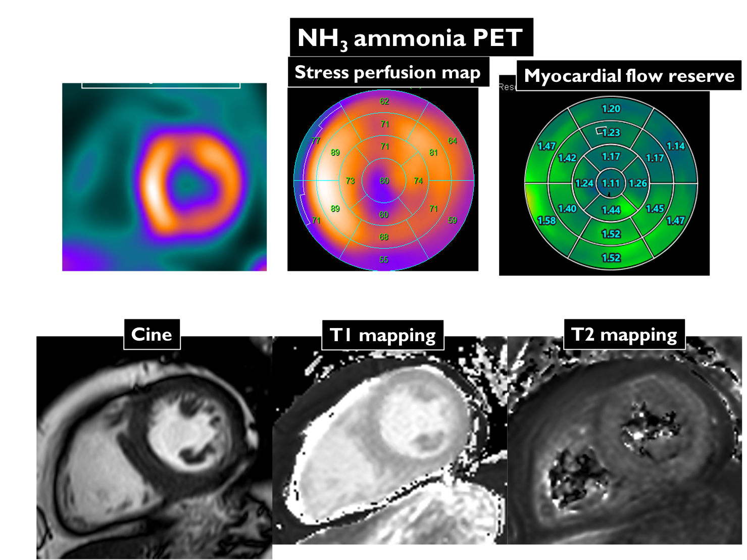
A man in his 60s man who underwent heart transplantation 16 years ago due to dilated cardiomyopathy. NH3-PET can quantify the absolute MBF and MFR by coronary territory (upper row). MRI T1 and T2 values in four territories were measured using the short-axis of mid left ventricular T1 and T2 mappings (lower row).

Comparison of T1 (left) and T2 (right) between territories with MFR >2.0 and <2.0.