Seonghwan Yee1, Lorna Browne1, and Justin Honce1
1Radiology, University of Colorado Anschutz Medical Campus, Aurora, CO, United States
1Radiology, University of Colorado Anschutz Medical Campus, Aurora, CO, United States
The MOLLI technique for T1 mapping was investigated using a phantom for its possible use as a T2-rho (T2 in the rotating frame) information tool. Since the T2rho is sensitive to iron content, the tool may be used as a composite relaxometry tool beyond the conventional use for cardiac T1 mapping.
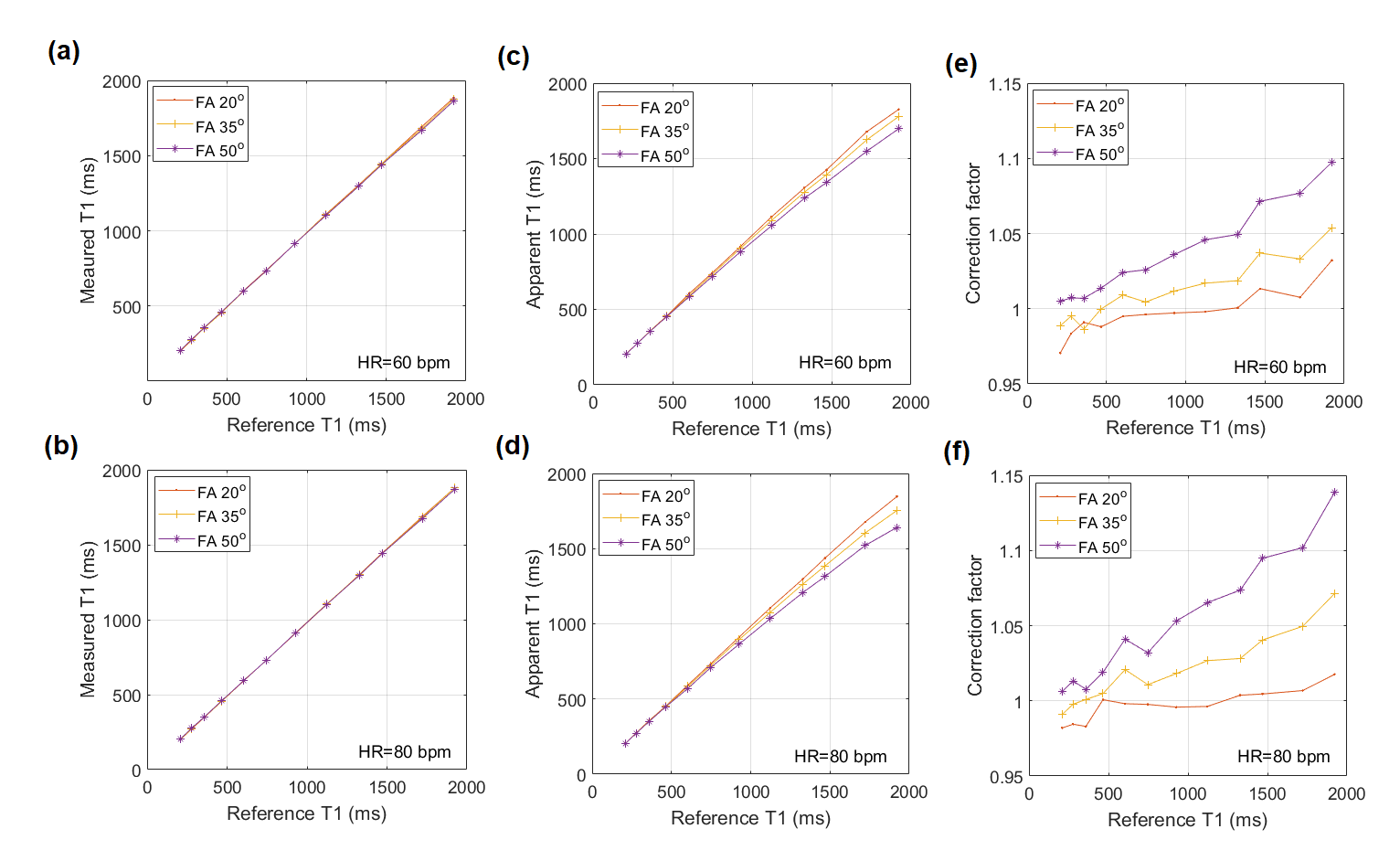
Fig. 4. In
comparison to the reference T1 values for the T1 phantom, the actual T1 values
measured by the MOLLI techniques are shown in (a) and (b). The top row is for
HR=60 bpm, while the bottom is for HR=80 bpm. The comparisons were made three
times for the flip angle (FA) 20°, 35° and 50°. The
apparent T1 comparisons for the three FAs are shown in (c) and (d), and the
correction factors are in (e) and (f).
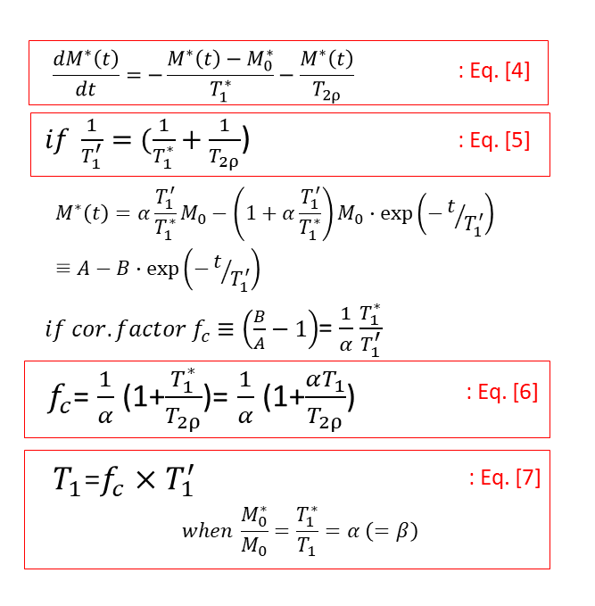
Fig. 2. If the
relaxation under the influence of the B1 field is non-negligible, the
longitudinal relaxation of the magnetization can be described as Eq. 4, where
the T2rho relaxation is added. If the apparent T1 relaxation, T1’ in Eq. 5, can
be expressed as Eq. 5, the signal model can still be the same way as Eq. 1 in
Fig.1, and the correction factor can be expressed as Eq. 6. The actual T1 can
still be obtained by multiplying the apparent T1 (T1’) and the correction
factor, as in Eq. 7.