Malte Roehl1,2, Peter D Gatehouse1,2, Pedro F Ferreira1,2, Sonya V Babu-Narayan1,2, David N Firmin1,2, Dudley J Pennell1,2, Sonia Nielles-Vallespin1,2, and Andrew D Scott1,2
1National Heart and Lung Institute, Imperial College London, London, United Kingdom, 2Cardiovascular Magnetic Resonance Unit, Royal Brompton Hospital, London, United Kingdom
1National Heart and Lung Institute, Imperial College London, London, United Kingdom, 2Cardiovascular Magnetic Resonance Unit, Royal Brompton Hospital, London, United Kingdom
To avoid errors in right ventricular T1 mapping caused by partial volume blood and epicardial fat signal we demonstrate a novel dark-blood fat-suppressed STEAM-SASHA method both in a phantom and in vivo.
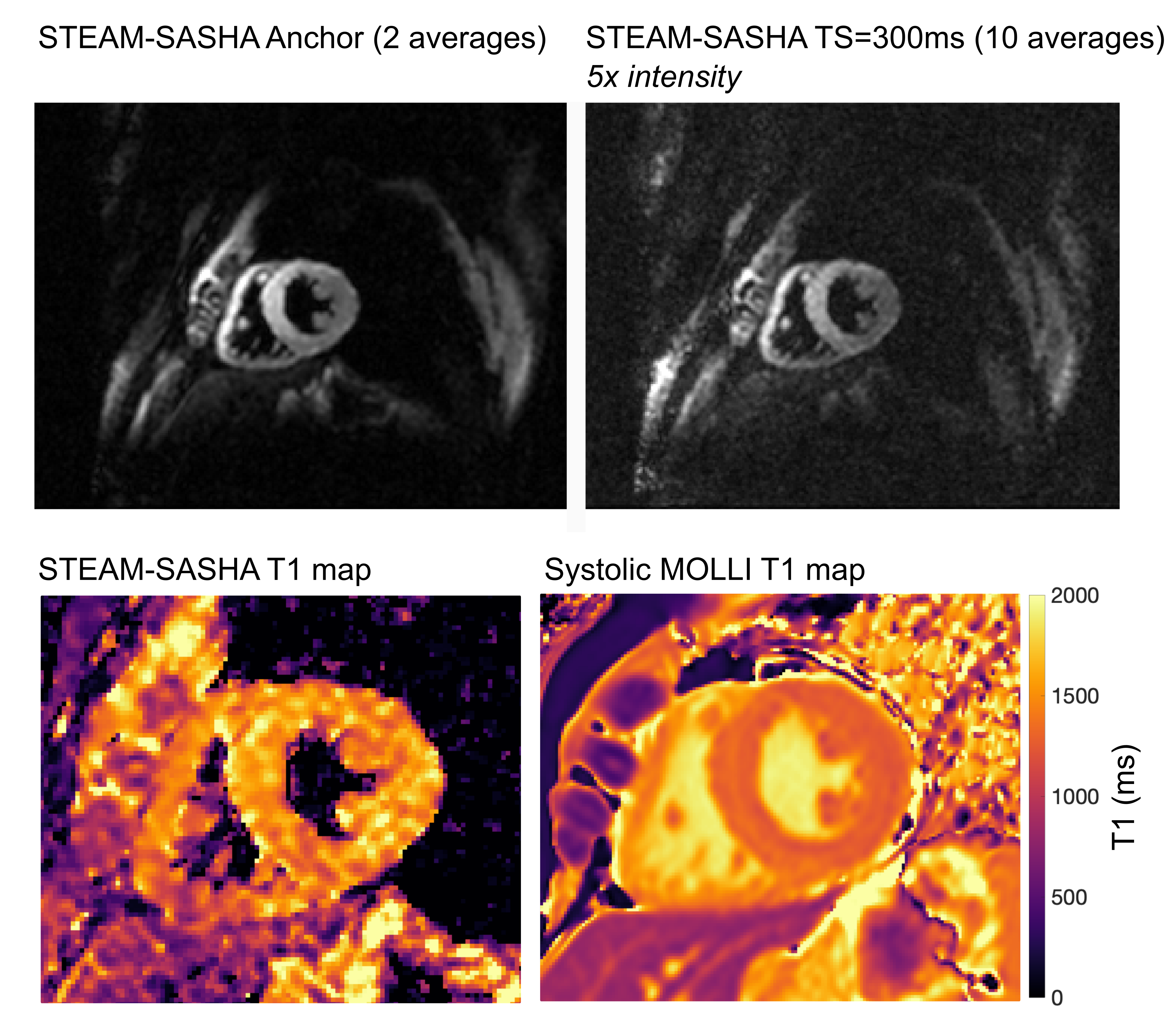
Figure 4:Full field of view example STEAM-SASHA images after
averaging two sequence repeats (top line) with the associated STEAM-SASHA T1 map
(bottom left) and the equivalent systolic product MOLLI T1 map acquired in one
typical volunteer (T1 maps cropped). The
STEAM-SASHA images show the reduction in signal intensity at the top and bottom
of the image due to the in-plane slice select gradient used. The STEAM-SASHA T1
map provides excellent blood and fat suppression in the left and right
ventricles.
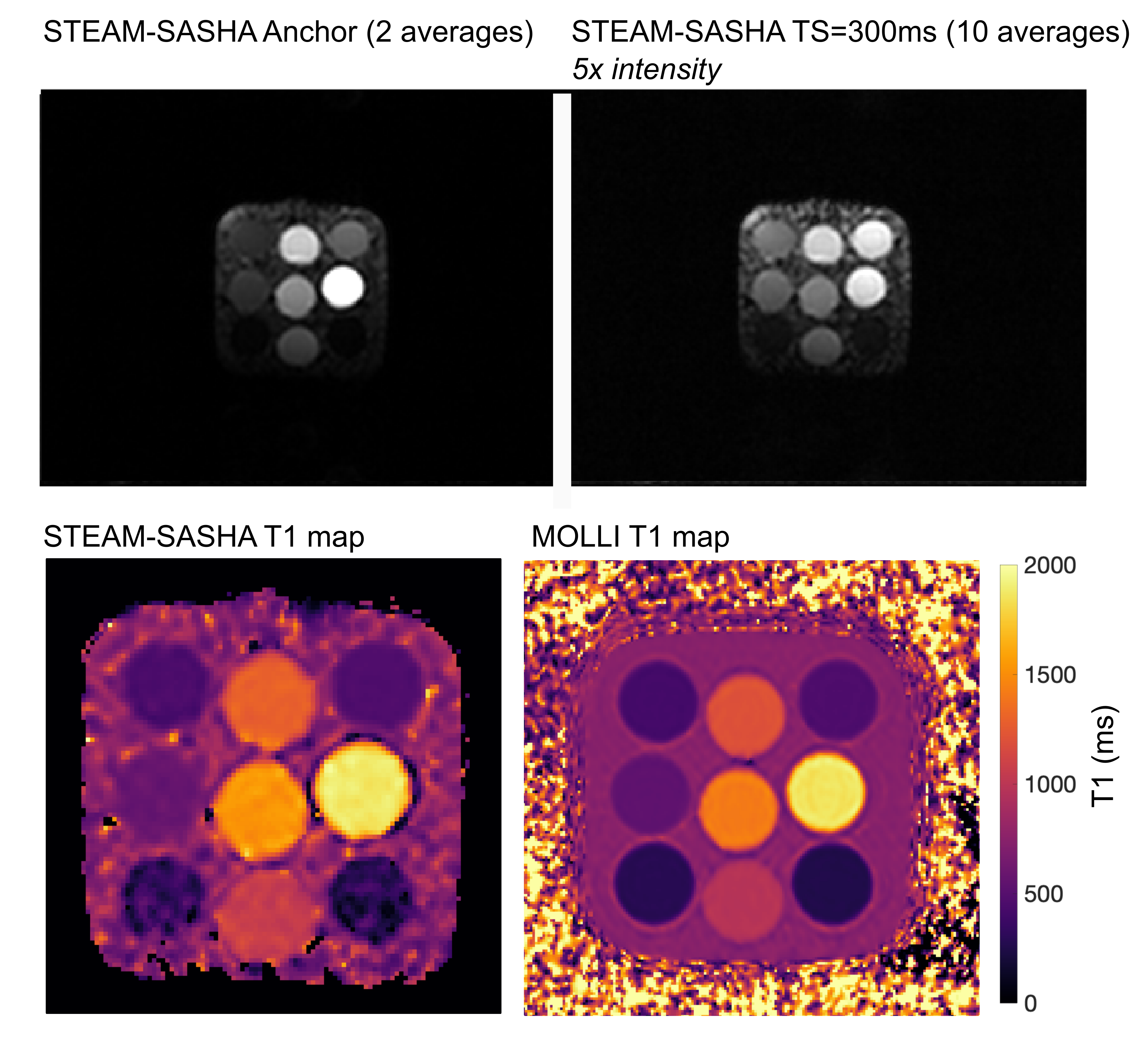
Figure 2:Example STEAM-SASHA images (full field of view) after
averaging two sequence repeats (top line) with the associated STEAM-SASHA map (cropped)
and the equivalent product MOLLI T1 map acquired in the T1MES phantom7.