Thibault Marin1, Paul K. Han1, Yue Zhuo1, Yanis Djebra1,2, Fang Liu1, Georges El Fakhri1, and Chao Ma1
1Massachusetts General Hospital, Harvard Medical School, Boston, MA, United States, 2LTCI, Telecom Paris, Institut Polytechnique de Paris, Paris, France
1Massachusetts General Hospital, Harvard Medical School, Boston, MA, United States, 2LTCI, Telecom Paris, Institut Polytechnique de Paris, Paris, France
Cardiac T1 mapping is a valuable tool to assess myocardial structure and assess cardiomyopathies. We develop a direct estimation method for estimation of 3D cardiac T1 mapping using subspace modeling and physical modeling though the Bloch equation.
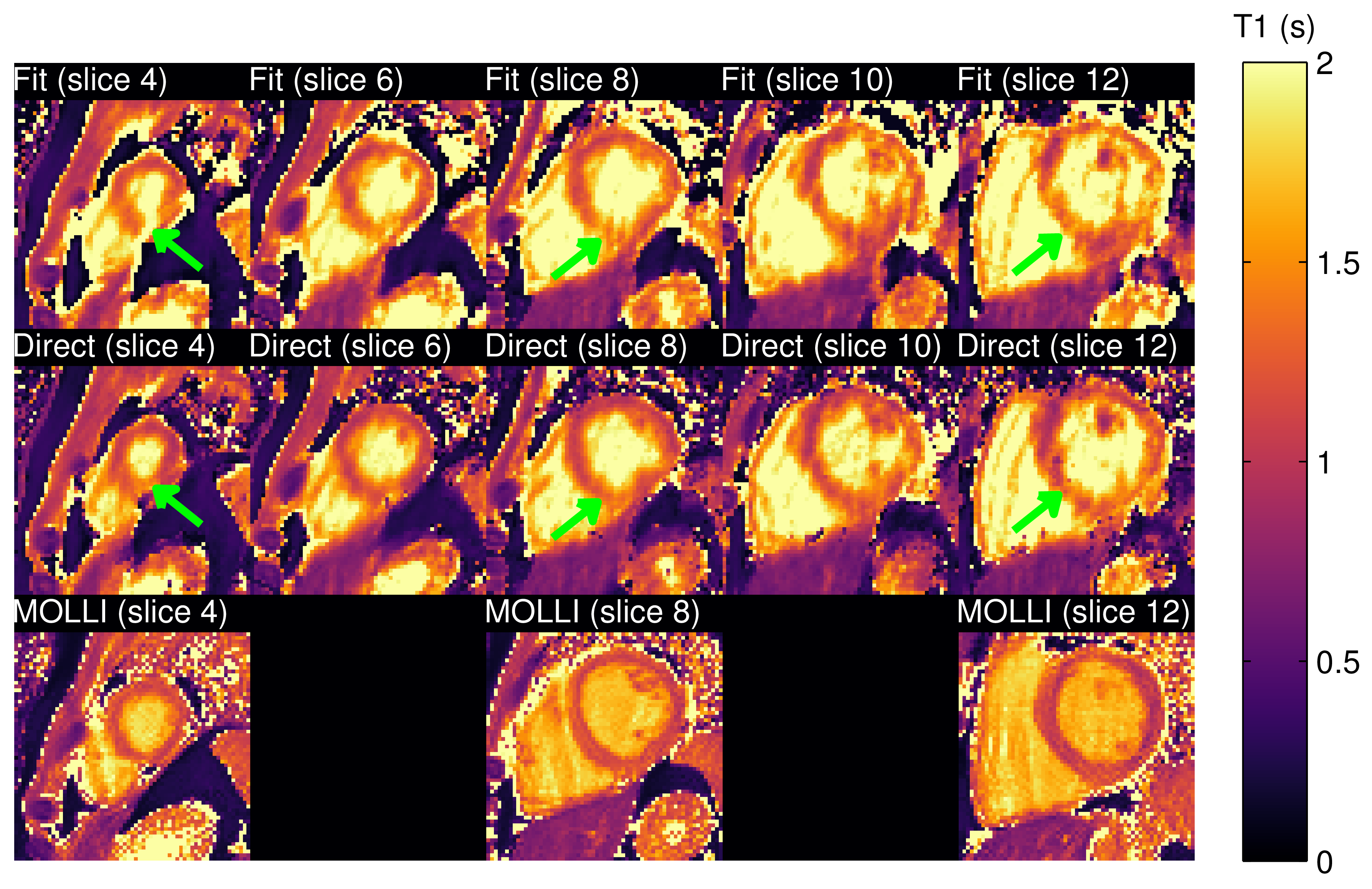
Figure 4. T1 mapping result. T1 maps obtained by simple
fitting of low-rank reconstruction (top row), the proposed direct
estimation method (middle row) and MOLLI (bottom row) are shown over
multiple slices. Green arrows indicate regions in the myocardium
where the Fit (top row) and Direct (middle row) differ. The T1 maps
from the proposed method better match those from MOLLI, with
correction of artifacts resulting in overestimation of T1 values
within the myocardium.
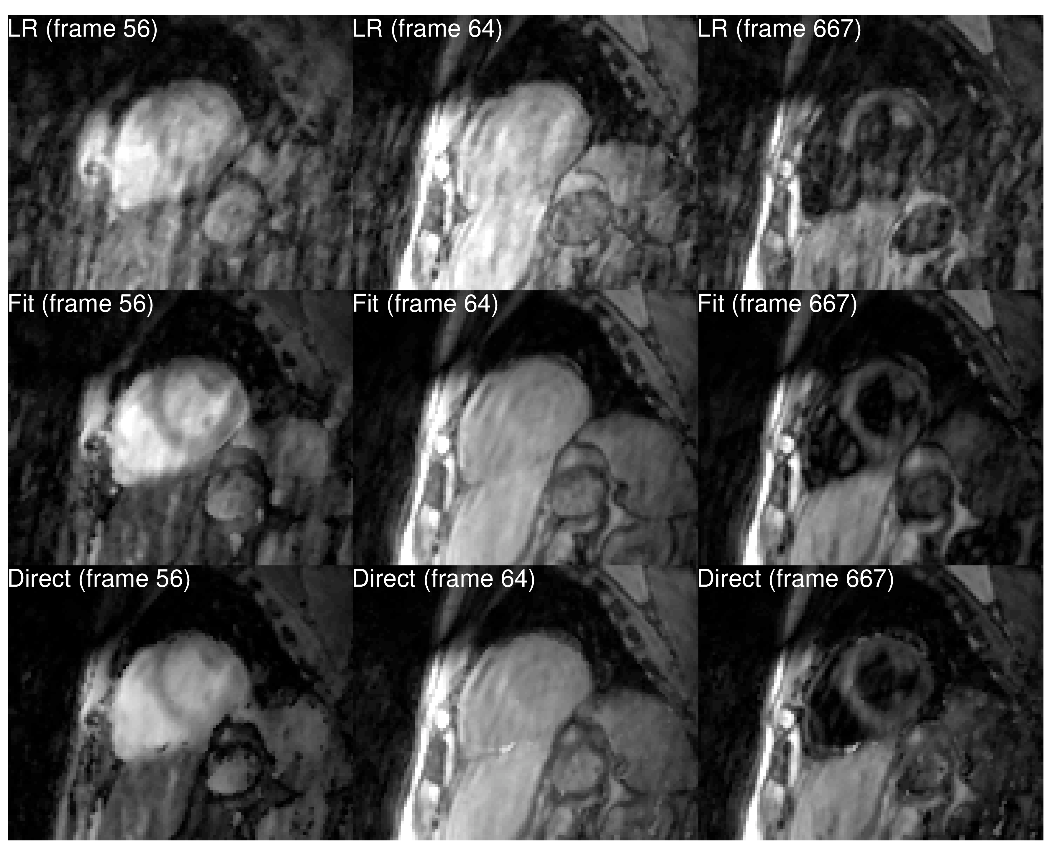
Figure 2. MR images at different time frames. Top-row
shows images from low-rank reconstruction (LR). The central row shows
synthesized images obtained from a T1 model fit of the low-rank
reconstruction (Fit). The bottom row shows temporal images obtained
using the proposed direct method (Direct). Low-rank reconstructions
are degraded by aliasing artifacts, which are reduced in synthesized
images obtained from T1 model fitting. The proposed method shows
improvement by further reducing the artifacts.