Daniel A Herzka1, Rajiv A Ramasawny1, Chris G. Bruce1, Delaney R. McGuirt1, William H. Schenke1, Jaffar M. Khan1, Adrienne E. Campbell-Washburn1,2, Aravindan A Kolandaivelu1,3, Toby A Rogers1,4, and Robert J. Lederman1
1NHLBI, Division of Intramural Research, National Institutes of Health, Bethesda, MD, United States, 2Biophysics and Biochemistry Branch, Division of Intramural Research, NHLBI, NIH, Bethesda, MD, United States, 3Division of Cardiology, Department of Medicine, Johns Hopkins University School of Medicine, Baltimore, MD, United States, 4Department of Cardiology, Medstar Washington Hospital Center, Washington, DC, United States
1NHLBI, Division of Intramural Research, National Institutes of Health, Bethesda, MD, United States, 2Biophysics and Biochemistry Branch, Division of Intramural Research, NHLBI, NIH, Bethesda, MD, United States, 3Division of Cardiology, Department of Medicine, Johns Hopkins University School of Medicine, Baltimore, MD, United States, 4Department of Cardiology, Medstar Washington Hospital Center, Washington, DC, United States
We examine 3D MRI to visualize chemoablation lesions created by intracoronary injection as used in septal ablation for the treatment of hypertrophic obstructive cardiomyopathy. In swine, 3D native contrast and gadolinium-enhanced MRI clearly delineated lesion extent.

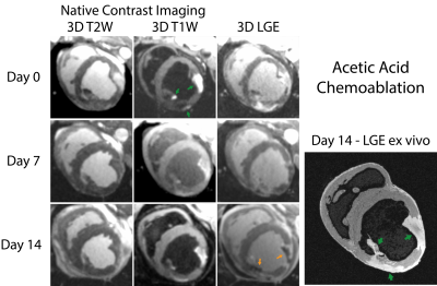
Figure 2: 3D imaging of acetic acid chemoablations (green arrows). Acutely, acetic acid produces strong T1 enhancement, which in combination with SPGR (inherently T1-weighted) results in signal contamination of T2W imaging. LGE appears hyperenhanced though heterogeneous. At later time points the T1W highlights lesion extent with good correspondence with ex-vivo MRI but with potentially confounding microvascular obstruction (orange arrows). Comparison with ex-vivo LGE indicates good correspondence to in-vivo imaging with both LGE and T1W imaging.

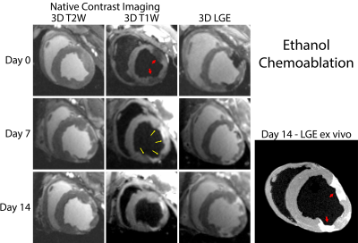
Figure 1: 3D imaging of ethanol chemoablation lesions (red arrows). Acutely, edema is mildly visible on T2-W imaging, T1-W imaging reflects hypointense signal, and lesions appear hypointense on LGE, indicating no contrast agent was present within lesions. At later time points the inherent T1 weighting of SPGR resulted in lesion visualization on T2-W imaging. 3D T1-W imaging resulted in hyperintense lesion cores, surrounded by thin layer of hypointensity depicting onset of encapsulating scar (yellow arrows). Comparison with ex-vivo imaging demonstrates good correspondence.
