Robert Jones1, Chiara Maffei1, Qiuyun Fan1, Jean Augustinack1, Barbara Wichtmann2, Aapo Nummenmaa1, Susie Huang1, and Anastasia Yendiki1
1Radiology, Athinoula A. Martinos Center for Biomedical Imaging, Massachusetts General Hospital and Harvard Medical School, Charlestown, MA, United States, 2Department of Radiology, University Hospital Bonn, Bonn, Germany, Bonn, Germany
1Radiology, Athinoula A. Martinos Center for Biomedical Imaging, Massachusetts General Hospital and Harvard Medical School, Charlestown, MA, United States, 2Department of Radiology, University Hospital Bonn, Bonn, Germany, Bonn, Germany
Ex vivo diffusion MRI signals exhibit bi-exponential decay as a function of q-value for both motor and association fibers. Parallel and perpendicular diffusion coefficients vary smoothly along each bundle, with marked differences between bundles.
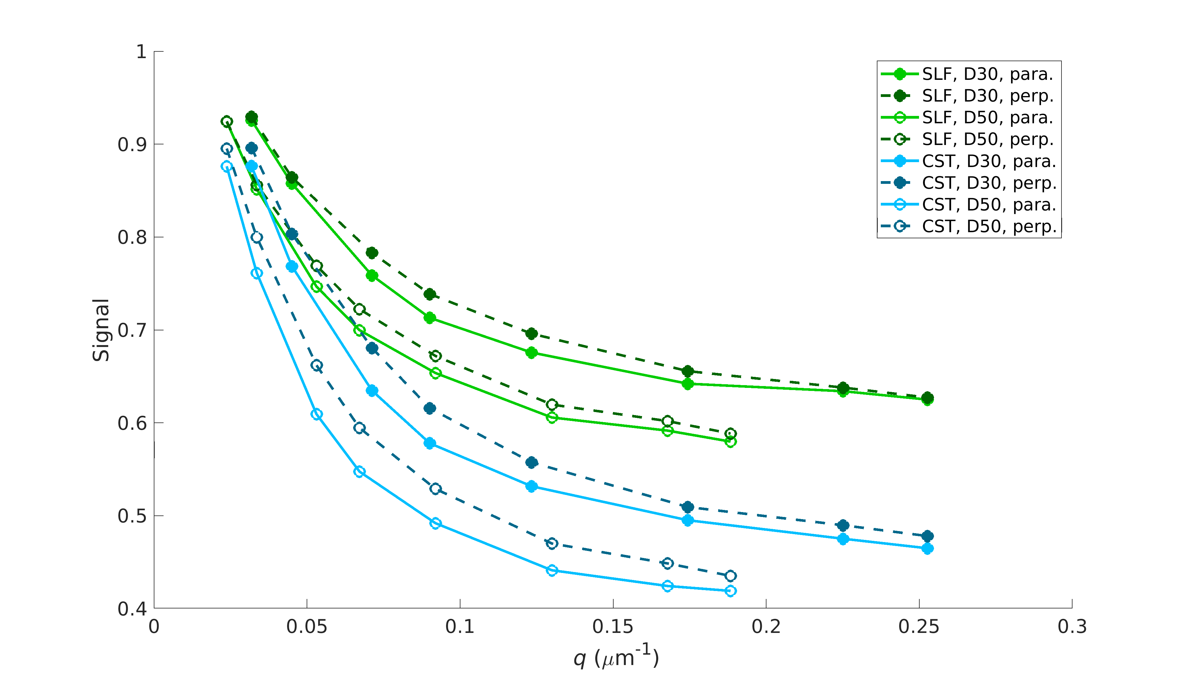
Figure 2. Decay curves for the diffusion signals parallel (light shade) and perpendicular (dark shade) to the fiber axis plotted as a function of q-value, for ROIs in the SLF (green) and CST (blue). Curves are shown for diffusion times of 30 ms (solid lines, filled markers) and 50 ms (dashed lines, open markers).
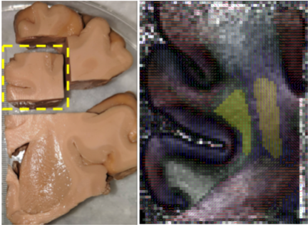
Figure 1. Left: Sample extracted from a coronal slab of a human hemisphere. Right: Locations of SLF (left) and CST (right) ROIs from one representative coronal slice, overlaid on the primary fiber orientation vectors and corresponding FA map from GQI reconstruction.