Ante Zhu1, J. Kevin DeMarco2,3, Robert Y. Shih2,3, Radhika Madhavan1, Tim Sprenger4, Chitresh Bhushan1, Maureen Hood2,3, Luca Marinelli1, Vincent B. Ho2,3, and Thomas K.F. Foo1
1GE Global Research, Niskayuna, NY, United States, 2Uniformed Services University of the Health Sciences, Bethesda, MD, United States, 3Walter Reed National Military Medical Center, Bethesda, MD, United States, 4GE Healthcare, Stockholm, Sweden
1GE Global Research, Niskayuna, NY, United States, 2Uniformed Services University of the Health Sciences, Bethesda, MD, United States, 3Walter Reed National Military Medical Center, Bethesda, MD, United States, 4GE Healthcare, Stockholm, Sweden
The axonal fiber parallel diffusivity (PD) measured by oscillating gradient spin echo is left-right symmetric in healthy corpus callosum. The genu and splenium showed regional difference of time-dependent PD, indicating different tissue tortuosity and randomly distributed restrictions.
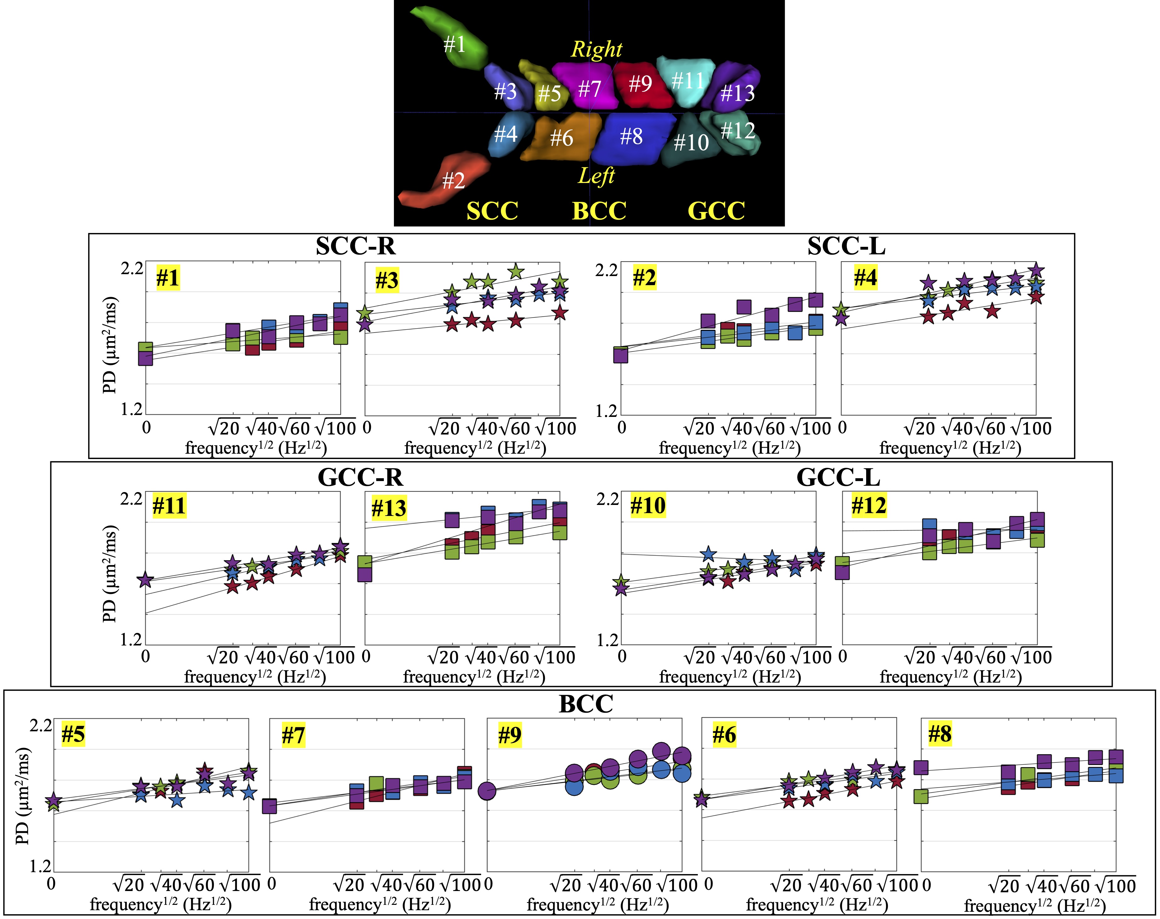
Figure 2. Segmented parcels #1-#13 of the corpus callosum; and parallel diffusivity measurements at different OGSE frequencies of the corpus callosum. Different colors represent different subjects. L: left; R: right.
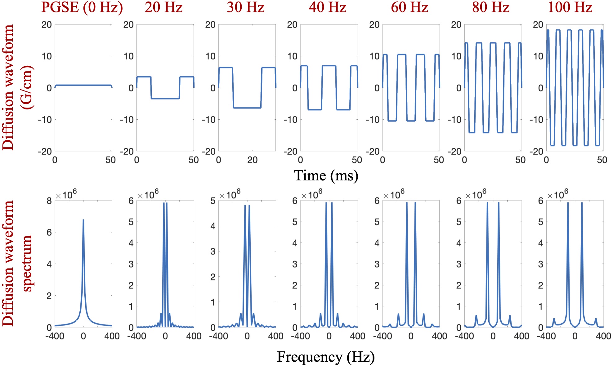
Figure 1. OGSE with different frequencies and PGSE with flattened waveforms used for diffusion imaging (top), and the corresponding diffusion spectrum (bottom).