Kevin B Borsos1,2, Desmond HY Tse2, Paul I Dubovan1,2, and Corey A Baron1,2,3
1Department of Medical Biophysics, Western University, London, ON, Canada, 2Centre for Functional and Metabolic Mapping, Western University, London, ON, Canada, 3Robarts Research Institute, Western University, London, ON, Canada
1Department of Medical Biophysics, Western University, London, ON, Canada, 2Centre for Functional and Metabolic Mapping, Western University, London, ON, Canada, 3Robarts Research Institute, Western University, London, ON, Canada
We present an optimized oscillating
gradient protocol to observe the difference in apparent kurtosis between OGSE
and PGSE acquisitions in vivo without
the requirement of a high-performance gradient insert.
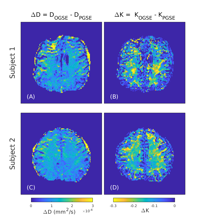
Figure 4: Differential ADC (A, C) and
differential kurtosis (B, D) maps for both subjects produced from the images shown in Figure 3. ΔD
and ΔK maps are generated from the subtraction of PGSE images from OGSE images
using ADC maps and apparent kurtosis maps respectively. The distortions in
the frontal lobe of Subject 1 are due to B0 inhomogeneity.
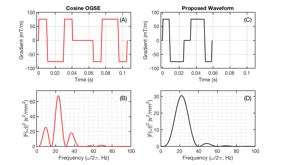
Figure 1: Gradient waveforms (A, C) and spectral densities (B, D)
of a conventional N = 2 cosine OGSE waveform (A) and our abbreviated oscillating
gradient waveform (C). Here the second diffusion gradient is inverted to reflect
the effect of the refocusing RF pulse (not shown). A reduction of the total
diffusion gradient duration is apparent for the new waveform (C) compared to
OGSE (A) at the same oscillation frequency of 25 Hz. Comparison of the
respective spectra in (B) and (D) shows similar spectral selectivity and
demonstrates zero DC spectral component for both waveforms.