Gregory Lemberskiy1, Santiago Coelho1, Thomas K.F. Foo2, Radhika Madhavan2, Luca Marinelli2, Jaemin Shin3, Els Fieremans1, and Dmitry S Novikov1
1Radiology, NYU School of Medicine, New York, NY, United States, 2GE Research, Niskayuna, NY, United States, 3GE Healthcare, New York, NY, United States
1Radiology, NYU School of Medicine, New York, NY, United States, 2GE Research, Niskayuna, NY, United States, 3GE Healthcare, New York, NY, United States
We present a near distortion-free and noise-free multishell diffusion neuro protocol at 1.5 mm enabled by the high performance MAGNUS head gradient coil reconstructed with random matrix theory denoising at the coil level. We showcase precise diffusion, kurtosis, and standard model maps.
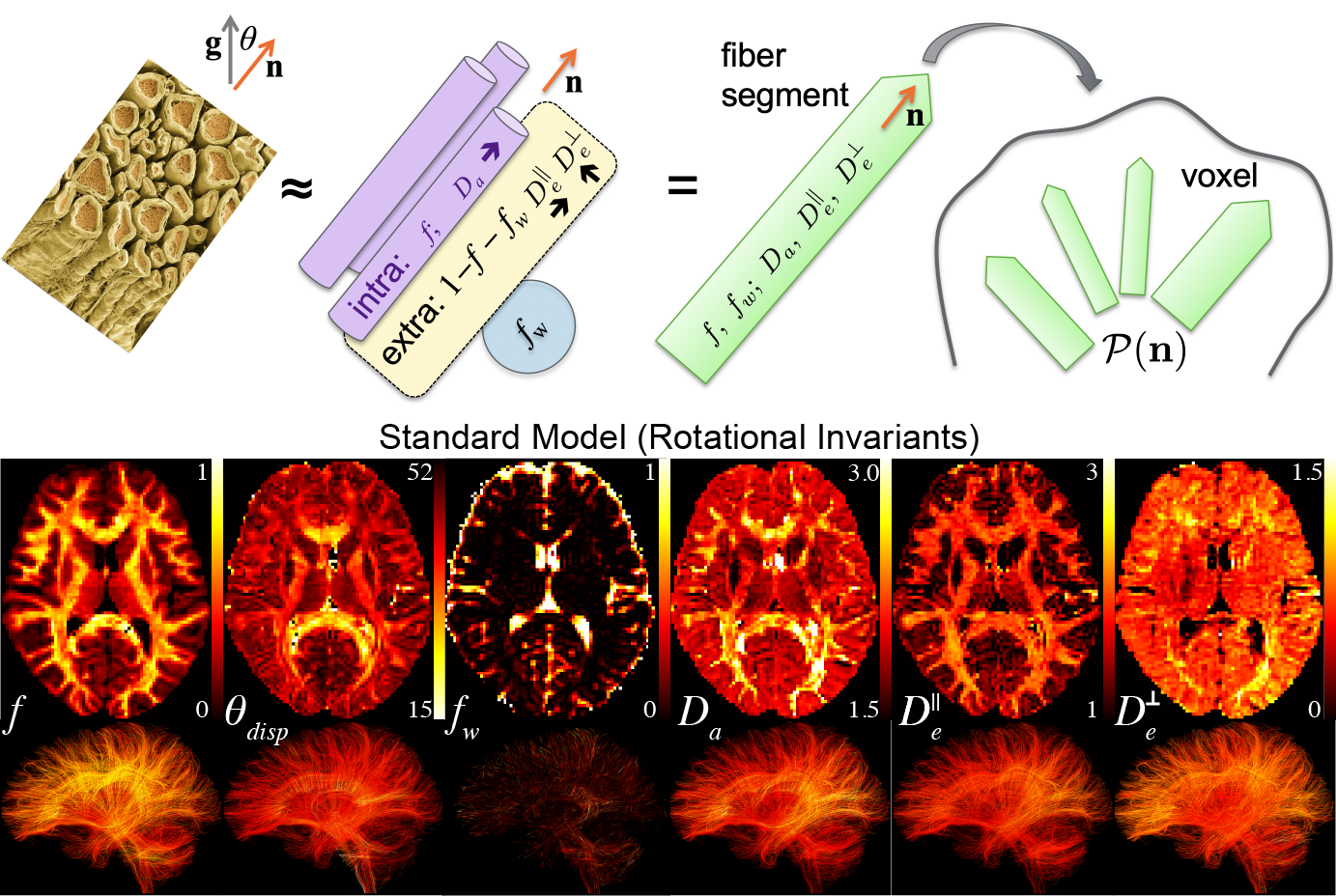
Standard Model. We show parametric maps for Standard Model parameters: axonal volume fraction $$$f$$$, free water fraction $$$f_w$$$, angular dispersion $$$\theta_{disp} = \cos^{-1}\sqrt{(2p_2+1)/3}$$$, axonal (neurite) diffusivity $$$D_a$$$, longitudinal extra-axonal diffusivity $$$D_e^\parallel$$$, and radial extra-axonal diffusivity $$$D_e^{\perp}$$$. Streamline tractograms are are colored by their correspsonding SM parameters.
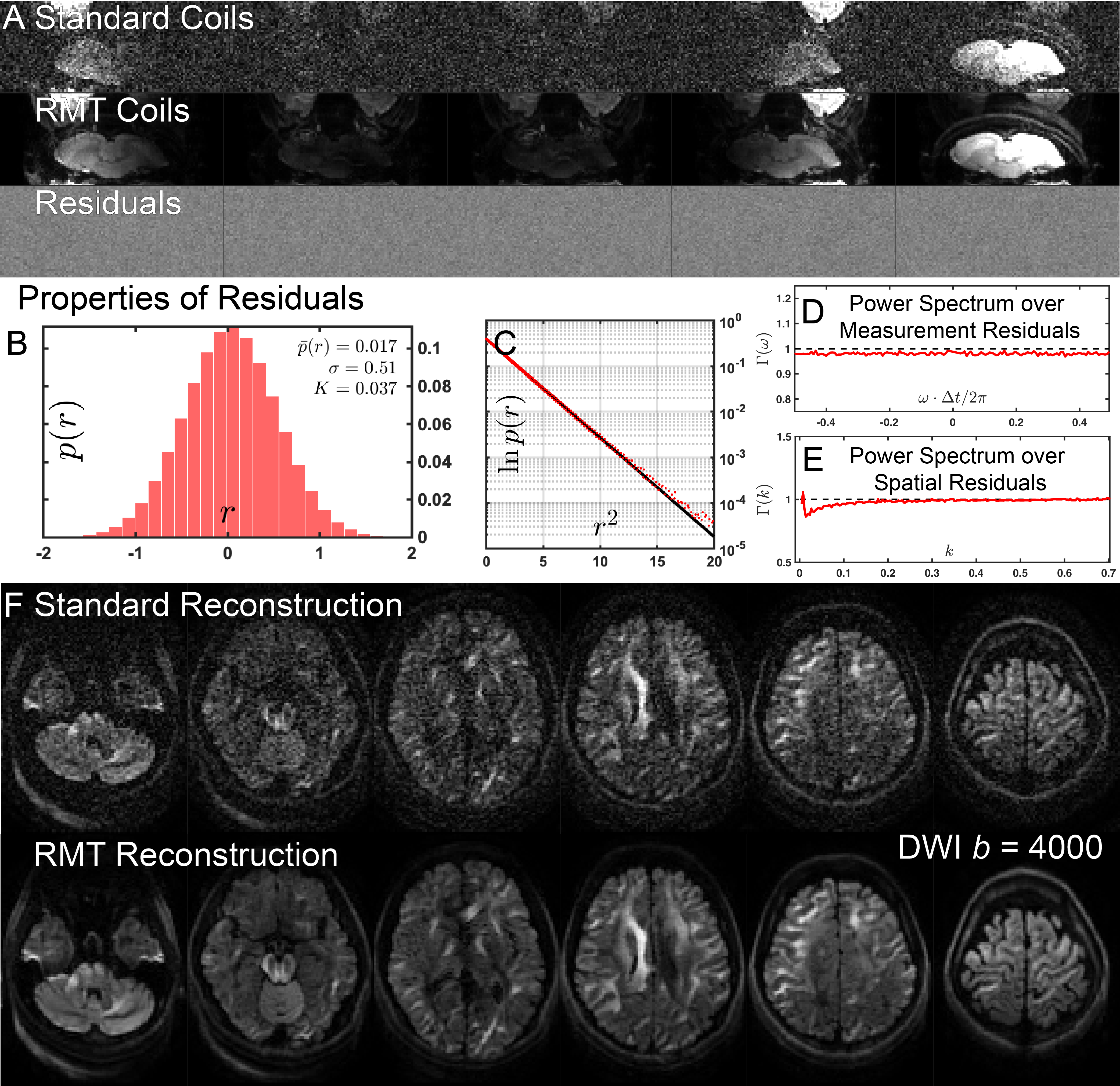
RMT Reconstruction. (A) 5/32 coils and their corresponding residuals are shown before and after noise removal via RMT. Properties of the removed normalized residuals $$$r$$$ are evaluated via (B,C) histograms $$$p(r)$$$, and (D,E) power spectrum analysis. (F) Standard and RMT reconstructions following parallel imaging and coil combination of a $$$b=4000\,$$$s/mm$$$^2$$$ image are displayed for several slices.