Zejun Wang1, Bao Wang2, Yingchao Liu3, and Ruiliang Bai1,4
1Key Laboratory of Biomedical Engineering of Ministry of Education, College of Biomedical Engineering and Instrument Science, Zhejiang University, Hangzhou, China, 2Department of Radiology, Qilu Hospital of Shandong University, Jinan, China, 3Department of Neurosurgery, Shandong Provincial Hospital Affiliated to Shandong First Medical University, Jinan, China, 4Department of Physical Medicine and Rehabilitation, Interdisciplinary Institute of Neuroscience and Technology, The Affiliated Sir Run Run Shaw Hospital, School of Medicine, Zhejiang University, Hangzhou, China
1Key Laboratory of Biomedical Engineering of Ministry of Education, College of Biomedical Engineering and Instrument Science, Zhejiang University, Hangzhou, China, 2Department of Radiology, Qilu Hospital of Shandong University, Jinan, China, 3Department of Neurosurgery, Shandong Provincial Hospital Affiliated to Shandong First Medical University, Jinan, China, 4Department of Physical Medicine and Rehabilitation, Interdisciplinary Institute of Neuroscience and Technology, The Affiliated Sir Run Run Shaw Hospital, School of Medicine, Zhejiang University, Hangzhou, China
We
compared shutter speed (SS) DCE-MRI and filter-exchange imaging (FEXI) for
vascular water exchange measurement in high-grade glioma. Our results
demonstrated consistent vascular water exchange assessments by SS
DCE-MRI and FEXI in both normal-appearing white matter and tumor.
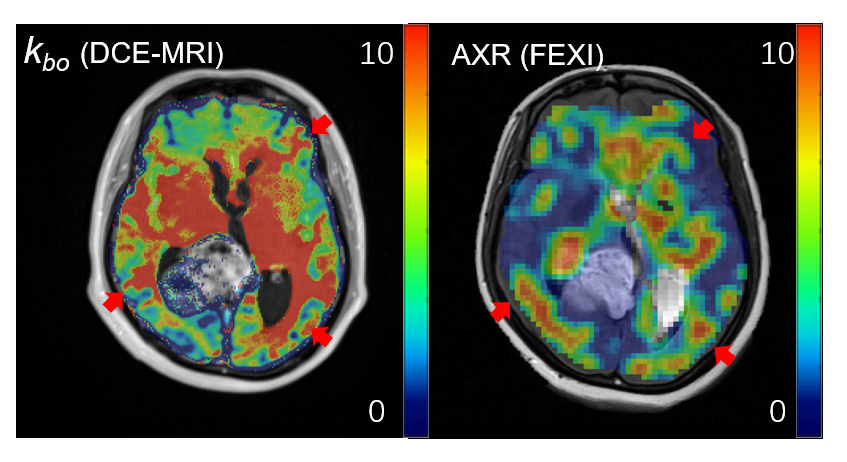
Figure 4: The
kbo map from
DCE-MRI and
AXR map from FEXI on the
same slice.
kbo’s
underlay is enhanced image, and AXR’s underlay is T2-weighted image. The kbo and AXR shows similar spatial patterns
as pointed with red arrows.
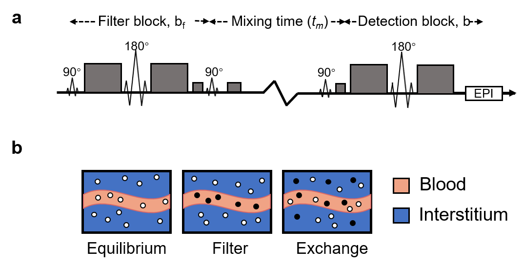
Figure
2: (a) The illustration of FEXI sequence. (b) At equilibrium, MR-visible water molecules (white dots) are located in blood with fast diffusivity
and interstitium with show diffusivity. Signal from fast diffusing intravascular water
is suppressed after filter applied, indicated by black dots,
leading to a
reduction in the ADC′. After tm, water
molecular exchange
leads to a recovery of
MR-visible water molecules in the
blood regions and the
ADC’ approaches the equilibrium ADC. Relaxation
back to the equilibrium ADC can be described by the apparent exchange rate
constant, AXR.