Samo Lasic1,2, Henrik Lundell1, Beata Wereszczyńska3, Matthew Budde4, Nadira Yuldasheva3, Filip Szczepankiewicz5, Erica Dall’Armellina3, Jürgen E. Schneider3, and Irvin Teh3
1Danish Research Centre for Magnetic Resonance, Centre for Functional and Diagnostic Imaging and Research, Copenhagen University Hospital Hvidovre, Copenhagen, Denmark, 2Random Walk Imaging, Lund, Sweden, 3Leeds Institute of Cardiovascular and Metabolic Medicine, University of Leeds, Leeds, United Kingdom, 4Department of Neurosurgery, Medical College of Wisconsin, Milwaukee, WI, United States, 5Clinical Sciences, Lund University, Lund, Sweden
1Danish Research Centre for Magnetic Resonance, Centre for Functional and Diagnostic Imaging and Research, Copenhagen University Hospital Hvidovre, Copenhagen, Denmark, 2Random Walk Imaging, Lund, Sweden, 3Leeds Institute of Cardiovascular and Metabolic Medicine, University of Leeds, Leeds, United Kingdom, 4Department of Neurosurgery, Medical College of Wisconsin, Milwaukee, WI, United States, 5Clinical Sciences, Lund University, Lund, Sweden
Acceleration compensated tensor-valued encoding at high b-values is feasible on a preclinical scanner. Time-dependent diffusion was observed in mouse hearts.
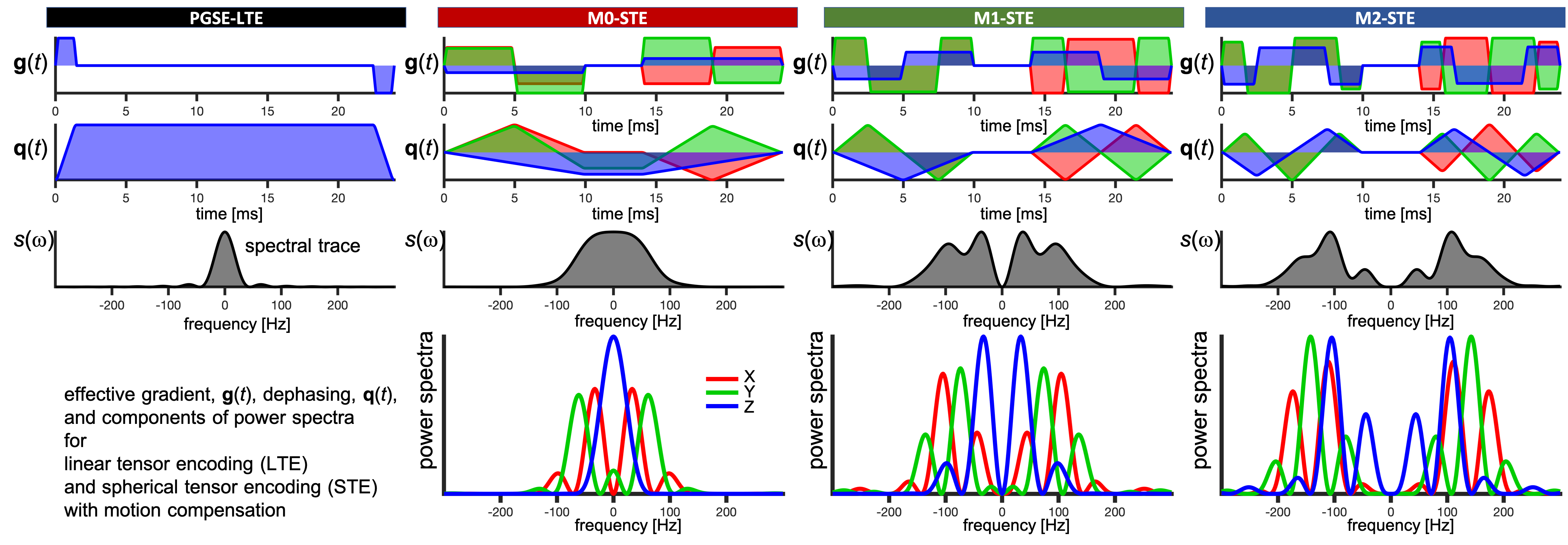
Figure 1: Effective gradient waveforms g(t), dephasing waveforms q(t), normalized spectral trace s(ω) and diagonal components of the dephasing cross power spectral density. Individual gradient components (XYZ shown in red, green, blue) from the motion compensated STE waveforms (M0-, M1-, M2-STE) were used as M0-, M1-, M2-LTE. with all moments nulled up to zeroth, first and second order (M0, M1, M2). Their power spectra is shown below s(ω). Note the shift of spectra from zero frequency to higher frequencies, which alters sensitivity to time-dependent diffusion.
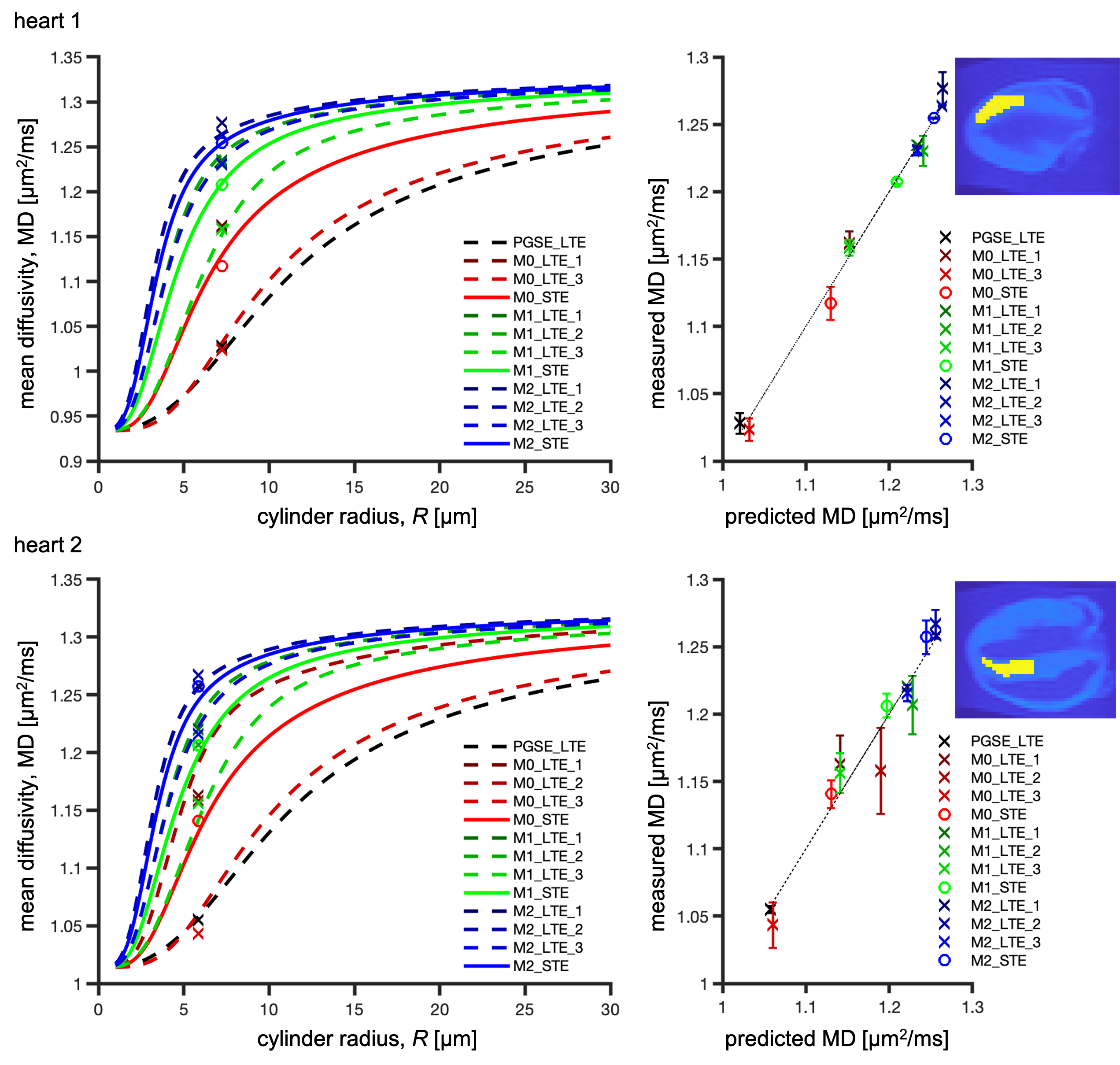
Figure 2: ROI-average mean diffusivity in the left ventricle myocardium in hearts 1 (top) and 2 (bottom) and theoretical prediction. The ROIs are shown the right. Left column: measured MD (markers) and prediction (lines) vs cylinder radius, R. Right column: measured MD vs prediction with error bars corresponding to the distance between measurement and prediction (dotted line for unity relation). The n=1,2,3 labels for Mn-LTE correspond to the X,Y,Z channels of Mn-STE.