Tokunori Kimura1, Kousuke Yamashita1, and Kouta Fukatsu1
1Department of Radiological Science, Shizuoka College of Medicalcare Science, Hamamatsu, Japan
1Department of Radiological Science, Shizuoka College of Medicalcare Science, Hamamatsu, Japan
We
clarified that our proposed T2wsup-DWI technique was superior to already
proposed water suppression DWI methods of FLAIR and non-b-zero (NZE) methods in
both of the ADC-SNR and the reduction effects of CSF partial volume effects
(PVE) in DTI parameters of ADC, FA, and fiber tractograpy.
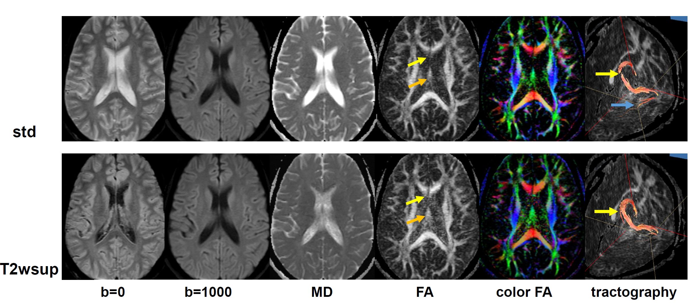
Fig. 4. Results of MR brain study Comparison of DTI
images, quantitative maps, and fiber tractographies, for standard (std) and for
our water suppression (T2wsup). The T2wsup provided better tissue-specific
values for the tissues close to the boundary regions of the ventricle, as the
body of fornix (orange arrows), or genu of corpus callosum (yellow arrows). For
the tractographies of fornix, the fibers at the central portions of two seed
ROIs (yellow arrow) for T2wsup was thicker and better connected than for the
standard, and artifactual fibers were drawn for the standard (blue arrow).
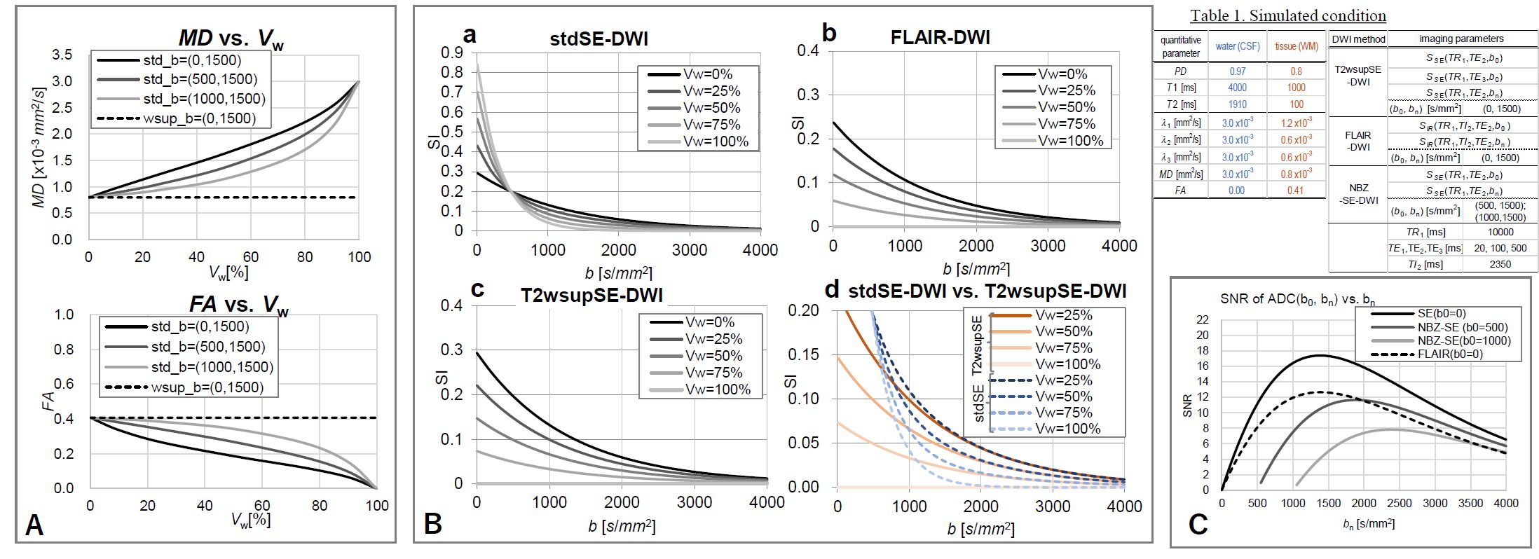
Fig. 3. Simulation results with 2-point
method. A: MD and FA as a function of Vw (%) for standard SE-DWI (std) and
T2wsupSE-DWI (T2wsup) obtained by the two-point method with denoted
combinations of b = (b0, bn) [s/mm2]. B: SIs as a function of b-value
(b) for three DWI methods (a–c) each as a parameter of water volume ratio (Vw),
and overlapped version a and b (d). C: Theoretical SNRs of ADC values as a
function of the second b-value, bn of pure tissue (Vw = 0,
ADC = 0.8×10⁻³ mm2/s) for three DWI techniques with two-point
method when the SNR of SE b0 image (S(b0)/σ) = 50.