Sajjad Feizollah1,2 and Christine L. Tardif1,2,3
1Department of Neurology and Neurosurgery, McGill University, Montreal, QC, Canada, 2McConnell Brain Imaging Center, Montreal Neurological Institute, McGill University, Montreal, QC, Canada, 3Department of Biomedical Engineering, McGill University, Montreal, QC, Canada
1Department of Neurology and Neurosurgery, McGill University, Montreal, QC, Canada, 2McConnell Brain Imaging Center, Montreal Neurological Institute, McGill University, Montreal, QC, Canada, 3Department of Biomedical Engineering, McGill University, Montreal, QC, Canada
A b-tensor diffusion imaging sequence with a spiral readout was
implemented at 7T with dynamic field monitoring. We present maps of the
microscopic anisotropy of the brain at 1.4 mm isotropic with minimal geometric
distortions.
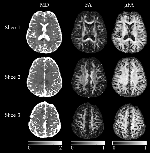
Figure 4- Mean
diffusivity (MD), fractional anisotropy (FA), and microscopic anisotropy (µFA) maps for 3 transverse slices.
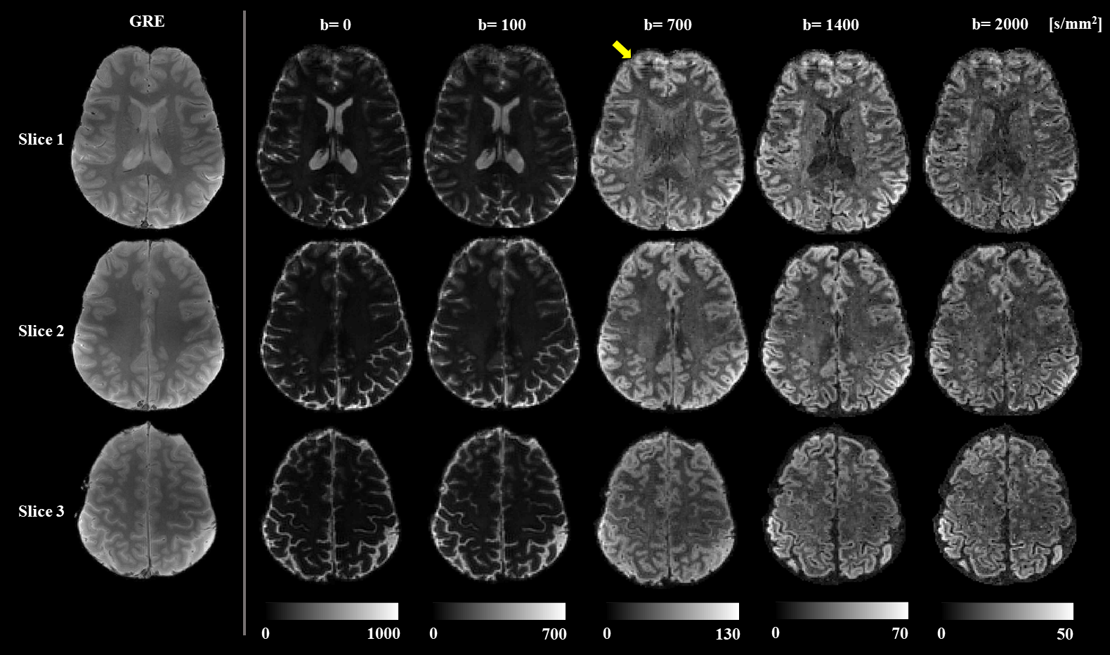
Figure 3-
Reconstructed images of 3 slices. The first column on the left shows images
acquired by the Cartesian GRE sequence for comparison. Other columns show
reconstructed STE images for 4 b-values with the same diffusion direction.
Images of each b-value are presented with a different scale for better
visibility. The yellow arrow shows remaining artifacts caused by static field inhomogeneities in the frontal lobe.