Nico J. J. Arezza1,2 and Corey A. Baron1,2
1Medical Biophysics, Western University, London, ON, Canada, 2Centre for Functional and Metabolic Mapping, Robarts Research Institute, London, ON, Canada
1Medical Biophysics, Western University, London, ON, Canada, 2Centre for Functional and Metabolic Mapping, Robarts Research Institute, London, ON, Canada
We investigated two techniques to remove free-water partial volume effects in microscopic fractional anisotropy (μFA) imaging. The techniques outperformed standard diffusion imaging methods in simulations and an expected increase in μFA and decrease in diffusivity were observed in vivo.
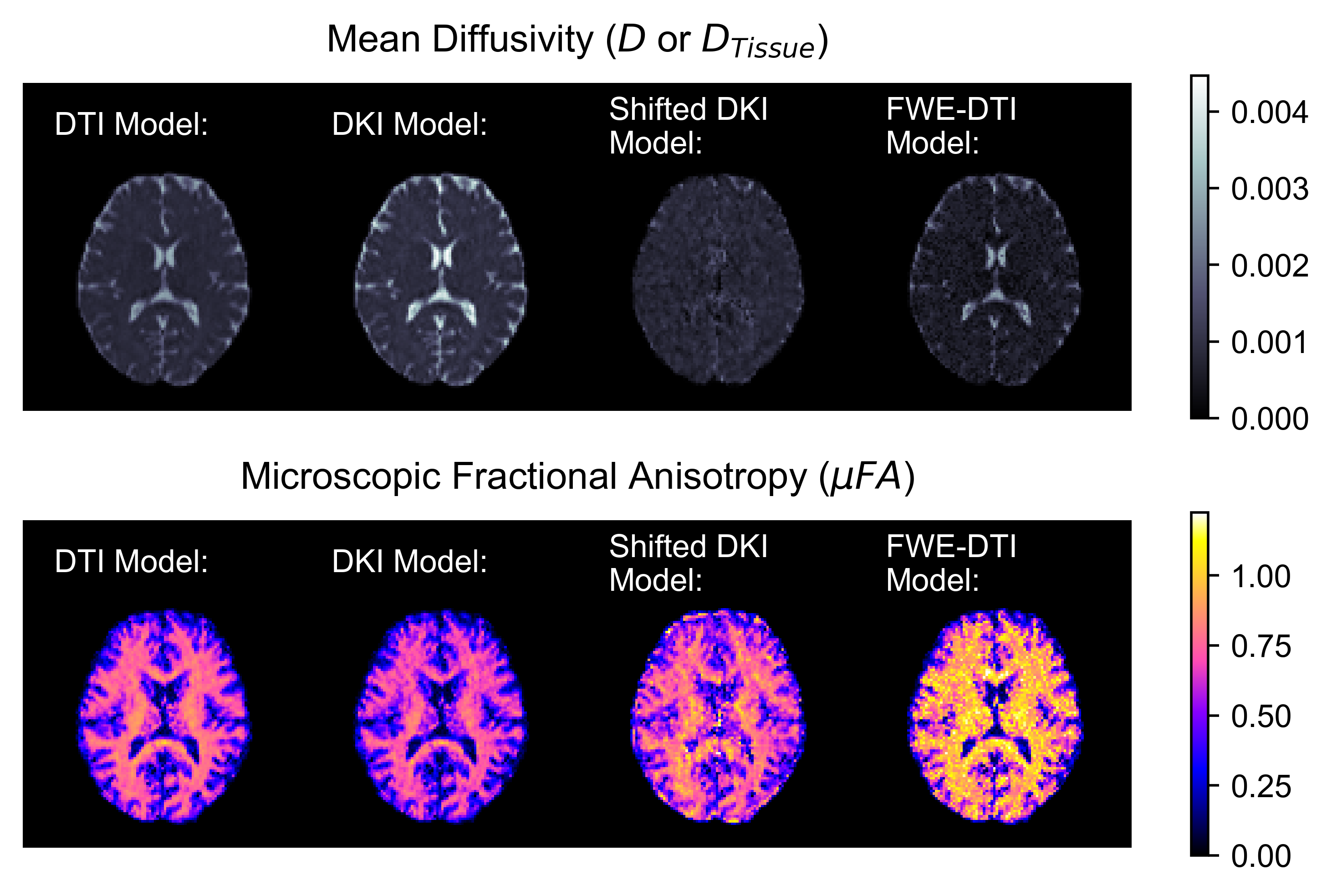
Figure 3. Example in vivo mean diffusivity (top) and microscopic fractional anisotropy (bottom) slices estimated using the DTI model (left), DKI model (center-left), shifted DKI model (center-right), and FWE-DTI model (right) with data from STE acquisitions.
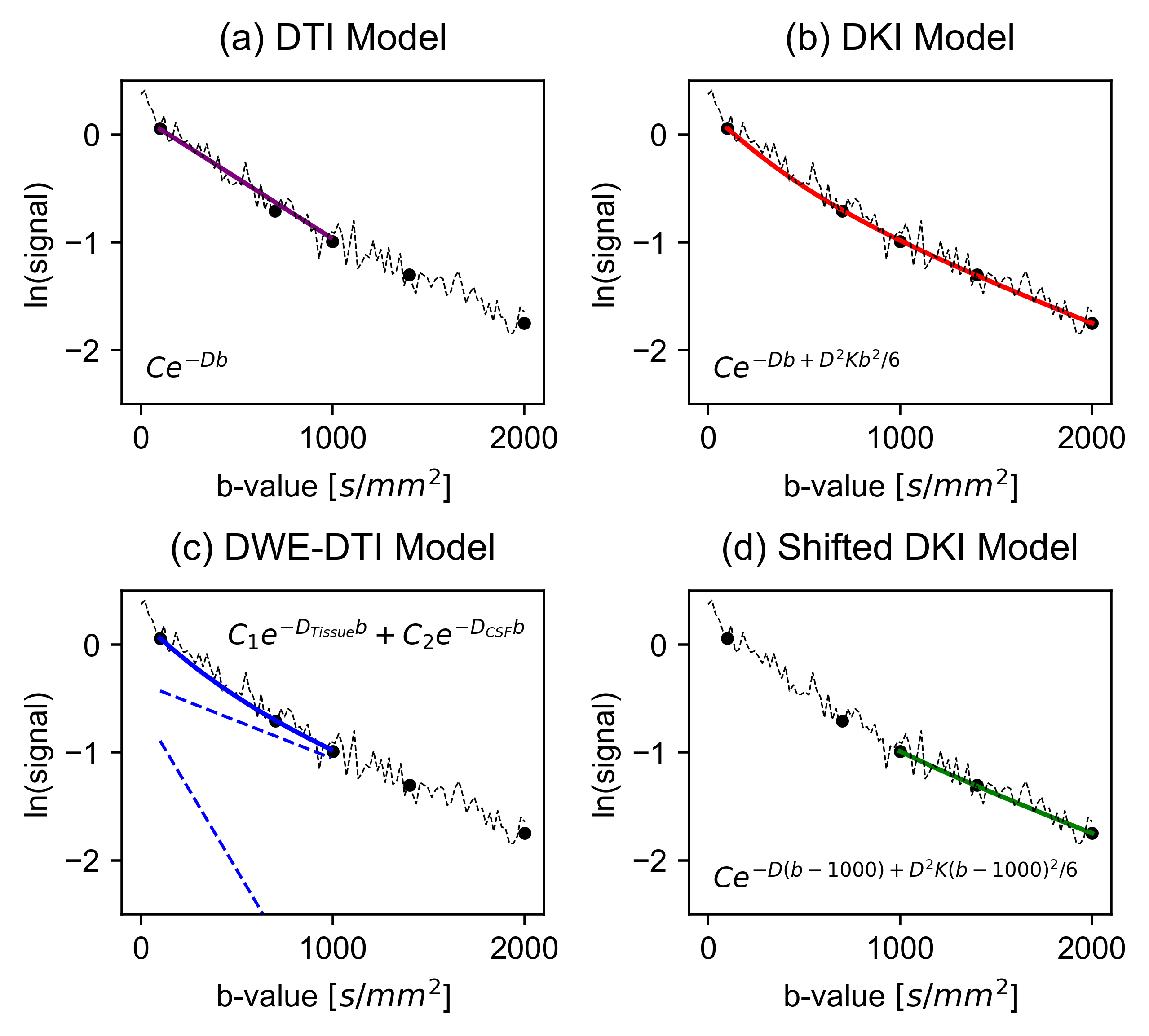
Figure 1. Summary of the models used to estimate diffusivity from STE signal data in voxels containing CSF and tissue: (a) DTI, (b) DKI, (c) FWE-DTI (with separate compartments shown by dashed lines), and (d) shifted DKI. The C terms represent constants. In FWE-DTI, signal due to CSF-PVEs is removed by separating the overall signal into diffusion components for free water and brain tissue. In shifted DKI, signal due to CSF contamination is attenuated via the use of higher b-values.