Cyrano Chatziantoniou1,2, Reineke Schoot2, Roelof van Ewijk2, Simone ter Horst2, Rick van Rijn3, Hans Merks2, Alexander Leemans1, and Alberto de Luca1
1Image Science Institute, UMC Utrecht, Utrecht, Netherlands, 2Princess Máxima Center for Pediatric Oncology, Utrecht, Netherlands, 3Department of Radiology, Academic Medical Centre Amsterdam, Amsterdam, Netherlands
1Image Science Institute, UMC Utrecht, Utrecht, Netherlands, 2Princess Máxima Center for Pediatric Oncology, Utrecht, Netherlands, 3Department of Radiology, Academic Medical Centre Amsterdam, Amsterdam, Netherlands
In surveying recent literature on sarcomas, we found that segmentation strategies vary strongly. In comparing these strategies on a toy example, we found that the choice of segmentation method can yield a difference in measured diffusion of up to 23%.
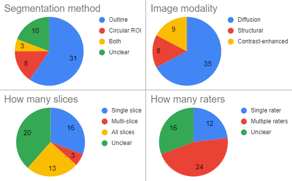
Figure 1: Segmentation methods as used in 52 recent papers on sarcoma shows a large variety in important aspects of the segmentation strategy.
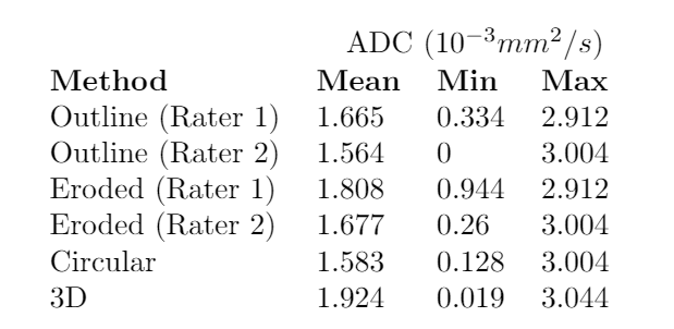
Choice of the segmentation strategy can have large impact on mean and minimum ADC (apparent diffusion coefficient) measured over the segmented area. Segmentation performed by two experienced radiologists (rater 1 and 2, outline) and a trained researcher (circular and 3D) on an ADC map of pediatric rhabdomyosarcoma. Segmented areas are shown in figure 3.