Archana Vadiraj Malagi1, Devasenathipathy Kandasamy2, Kedar Khare3, Deepam Pushpam4, Rakesh Kumar5, Sameer Bakhshi4, and Amit Mehndiratta1,6
1Centre for Biomedical Engineering, Indian Institute of Technology Delhi, New Delhi, India, 2Department of Radiodiagnosis, All India Institute of Medical Sciences Delhi, New Delhi, India, 3Department of Physics, Indian Institute of Technology Delhi, New Delhi, India, 4Department of Medical Oncology, Dr. B.R. Ambedkar Institute-Rotary Cancer Hospital (IRCH), All India Institute of Medical Sciences Delhi, New Delhi, India, 5Department of Nuclear Medicine, All India Institute of Medical Sciences Delhi, New Delhi, India, 6Department of Biomedical Engineering, All India Institute of Medical Sciences Delhi, New Delhi, India
1Centre for Biomedical Engineering, Indian Institute of Technology Delhi, New Delhi, India, 2Department of Radiodiagnosis, All India Institute of Medical Sciences Delhi, New Delhi, India, 3Department of Physics, Indian Institute of Technology Delhi, New Delhi, India, 4Department of Medical Oncology, Dr. B.R. Ambedkar Institute-Rotary Cancer Hospital (IRCH), All India Institute of Medical Sciences Delhi, New Delhi, India, 5Department of Nuclear Medicine, All India Institute of Medical Sciences Delhi, New Delhi, India, 6Department of Biomedical Engineering, All India Institute of Medical Sciences Delhi, New Delhi, India
Correlation analysis showed no significant
relationship between IVIM-DKI and PET parameters. IVIM parameters with Total-Variation
model produced substantial reproducibility with better quality parameter maps.
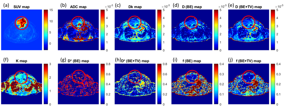
Figure2 (a) SUV map, (b) ADC map, (c, k) DKI parameter maps, (d, e) diffusion parameters
and (g-j) perfusion parameters from IVIM analysis of a 32-year-old male was
diagnosed for NHL; the tumour was present in sternum encircled in red.
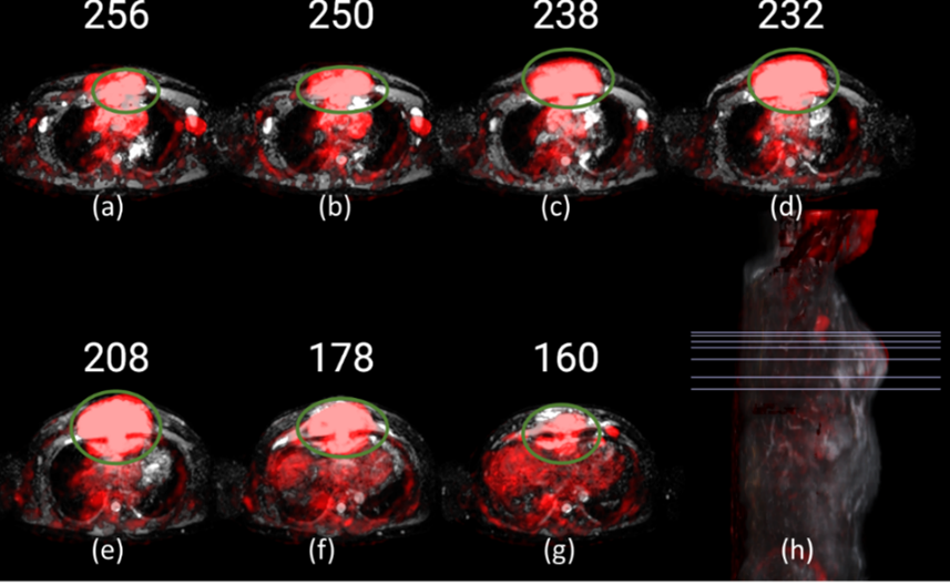
Figure1 PET images (red) overlapped onto ADC images of a patient with NHL, where the tumour is encircled (green). (a) to (g) represents
baseline scans at axial view. (h) Representative 3D sagittal view with lines
indicating slices (a-g) selected for visualization.