Amaresha Shridhar Konar1, Akash Deelip Shah2, Ramesh Paudyal1, Jung Hun Oh1, Eve LoCastro1, David Aramburu Nuñez1, Nathaniel Swinburne2, Robert J. Young2, Andrei I. Holodny2, Kathryn Beal3, Vaios Hatzoglou2, and Amita Shukla-Dave1,2
1Medical Physics, Memorial Sloan Kettering Cancer Center, New York, NY, United States, 2Radiology, Memorial Sloan Kettering Cancer Center, New York, NY, United States, 3Radiation Oncology, Memorial Sloan Kettering Cancer Center, New York, NY, United States
1Medical Physics, Memorial Sloan Kettering Cancer Center, New York, NY, United States, 2Radiology, Memorial Sloan Kettering Cancer Center, New York, NY, United States, 3Radiation Oncology, Memorial Sloan Kettering Cancer Center, New York, NY, United States
The present
prospective study aims to determine the ability of DW- and DCE-MRI to predict the long-term response of Brain Metastases
within 72 hours of SRS. The results are promising as it will inform
the treating physicians at an early time point about which patients will
benefit from SRS (or not).
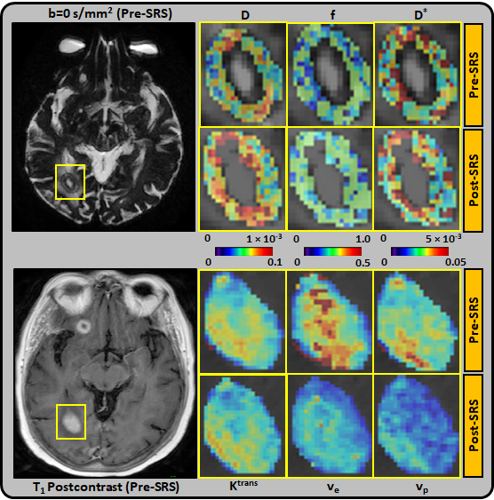
Figure 1: Representative pre-SRS and post-SRS (within 72 hours of treatment)
MR images from an 80 year-old female with right occipital lobe brain
metastasis, which demonstrated progressive disease (PD). Top panel: D(×10-3 mm2/s), D*(×10-3 mm2/s), and f maps are zoomed in at the
locations of ROIs (pre-SRS b=0 s/mm2 image). The lesion volume was
unchanged at post-SRS. Bottom panel: Ktrans (min-1), ve, and
vp maps are zoomed in at the locations of ROIs (pre-SRS T1-postcontrast
image).
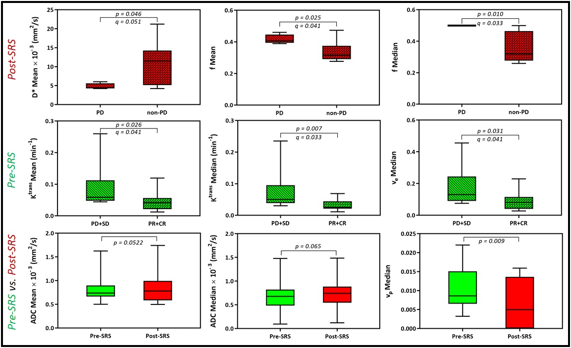
Figure 4: Lesions that progressed had a higher f mean and median than those
that did not. Lesions that responded to SRS had a lower pre-SRS Ktrans
mean, Ktrans median, and ve median than those that did
not. ADC mean and median trended higher in lesions post-SRS than pre-SRS. vp
was lower in lesions post-SRS than pre-SRS. ADC, apparent diffusion
coefficient. Ktrans, index of tumor vascular permeability. ve,
volume fraction of extracellular extravascular space. vp, vascular
volume.