ruicheng ba1, Hongxi Zhang2, Zhongwei Gu3, Yuhao Liao1, Xingwang Yong1, Zhiyong Zhao1, Yi Zhang1, and Dan Wu1
1Key Laboratory for Biomedical Engineering of Ministry of Education, Department of Biomedical Engineering, College of Biomedical Engineering & Instrument Science, Zhejiang University, Hang zhou, Zhejiang, China, 2Children's Hospital, Zhejiang University School of Medicne,Department of Radiology, Hang zhou, Zhejiang, China, 3Children's Hospital, Zhejiang University School of Medicne, Department of Pathology, Hangzhou, Zhejiang, China
1Key Laboratory for Biomedical Engineering of Ministry of Education, Department of Biomedical Engineering, College of Biomedical Engineering & Instrument Science, Zhejiang University, Hang zhou, Zhejiang, China, 2Children's Hospital, Zhejiang University School of Medicne,Department of Radiology, Hang zhou, Zhejiang, China, 3Children's Hospital, Zhejiang University School of Medicne, Department of Pathology, Hangzhou, Zhejiang, China
Tumor microstructure was estimated based on
time-dependent dMRI and IMPULSED model. Cellularity and intracellular
fraction increased from low-grade to high-grade glioma, and further increased
in medulobastom. Cellularity achieved the highest area-under-the-curve in
grading glioma.
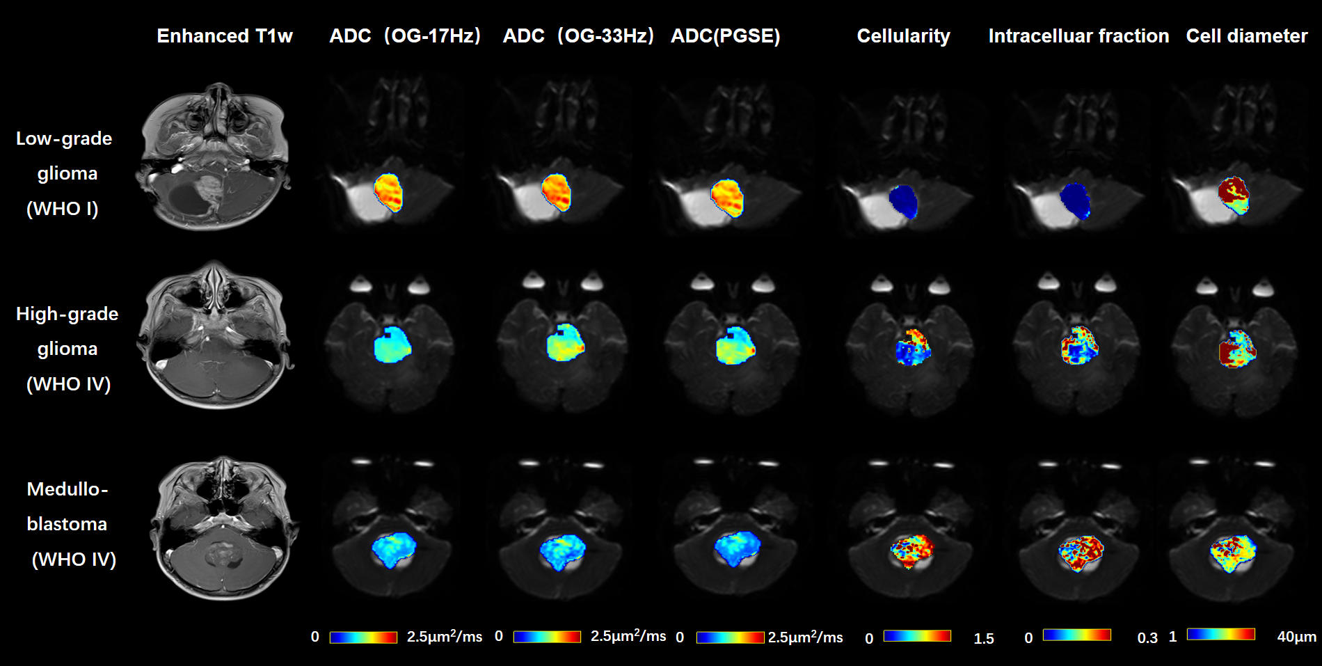
Figure 1: Gd-enhanced T1w images
and the maps of ADC(OGSE-17Hz, OGSE-33Hz, PGSE), cellularity,
intracelluar fraction and cell diameter as obtained from the IMPULSED model.
The
ADC and microstructural maps were overlaid on the b0 image for reprehensive
low-grade glioma, high-grade glioma, and medulloblastoma cases.
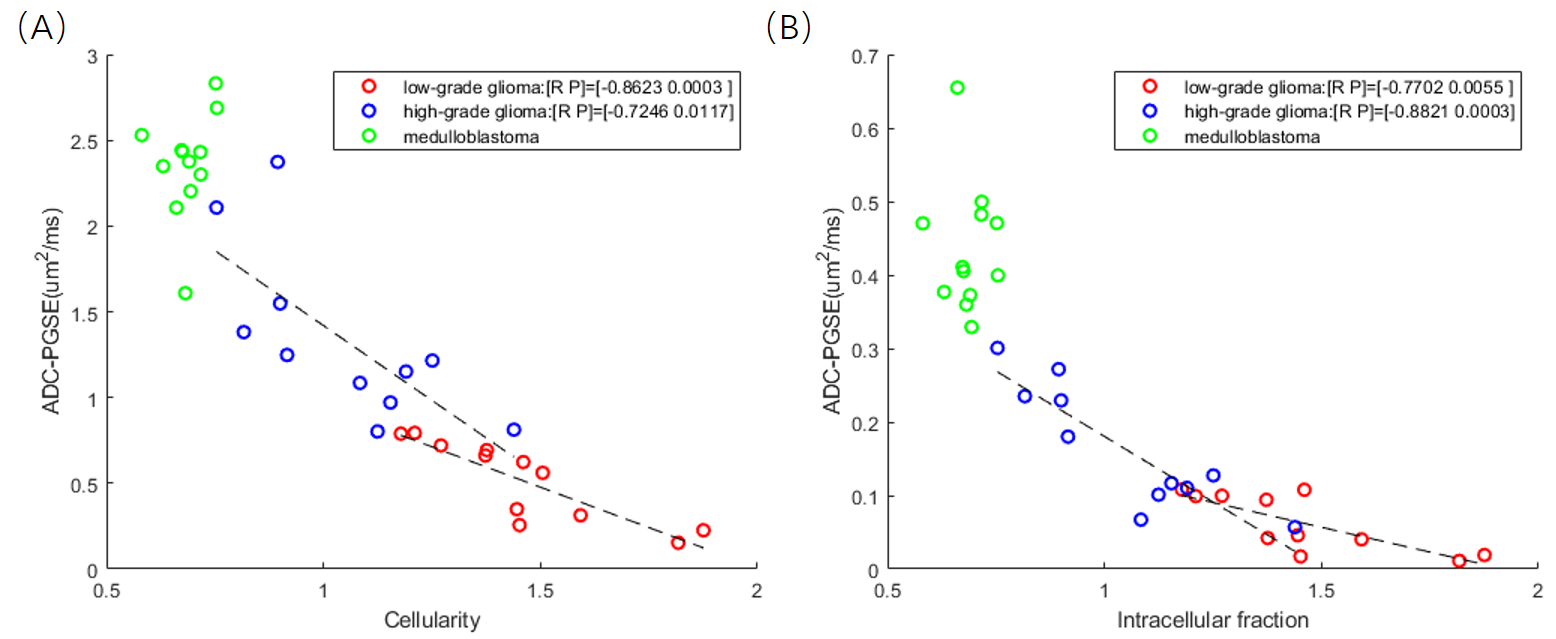
Figure 4: (A) Correlation between ADC (PGSE)
and cellularity. Regression analysis revealed correlation was strongest for low-grade glioma (r = -0.8623, p = 0.0003), followed by high-grade
glioma (r = -0.7246, p = 0.0117); whereas not significant correlation were
found for medulloblastoma (p=0.4095).
(B) Similar correlations patterns were found between ADC (PGSE) and Intracellular
fraction.