Rifeng Jiang1, Kaiji Deng1, Yixin Guo2, and Zhongshuai Zhang3
1Fujian Medical University Union Hospital, Fuzhou, China, 2Fujian Medical University, Fuzhou, China, 3MR Scientific Marketing, Siemens Healthcare, Shanghai, China
1Fujian Medical University Union Hospital, Fuzhou, China, 2Fujian Medical University, Fuzhou, China, 3MR Scientific Marketing, Siemens Healthcare, Shanghai, China
This study found that NODDI seems to be a more potent approach in evaluating the early CST infiltration by HGG, and can evaluate the CST destruction with a similar performance to MD by providing additional information about neurite density for HGG-induced CST injury.
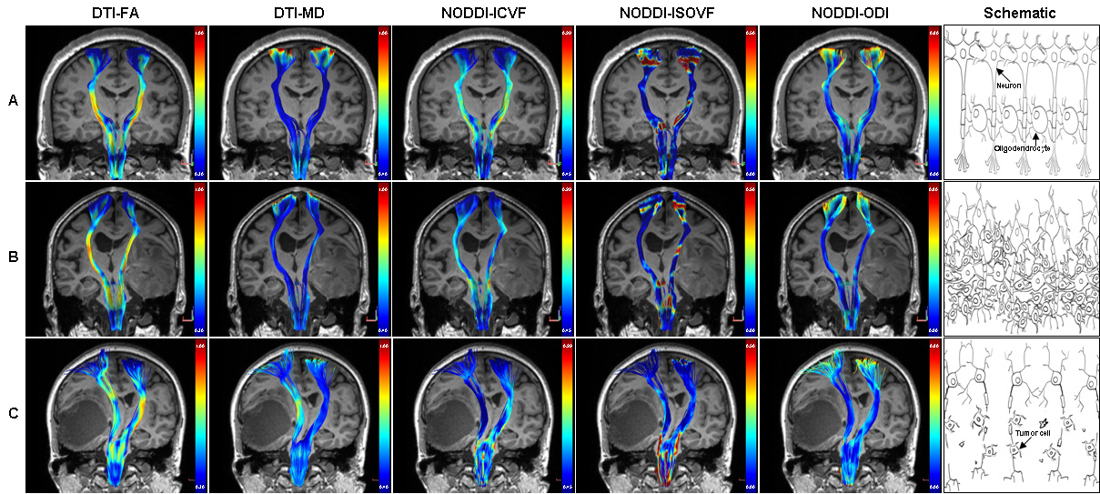
Three representative subjects demonstrating the CST
changes in HGG patients. The
subject A was a 39-year-old healthy male. All the CST features have good bilateral
symmetry. The patient B was a 72-year-old male with glioblastoma,
and his motor function was normal. The ICVF of the affected CST decreased
obviously, but changes of the affected CST were not obvious in the other
diffusion parameters. The patient C was a 62-year-old female with glioblastoma, and she had motor weakness (grade 2). Changes of the affected
CST were obvious in FA, MD and ICVF, but not in ISOVF and ODI.
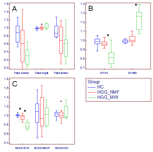
Changes of relative CST features in HGG patients. Box and whisker plot (A-C) showed that compared with the relative CST features in HGG_NMF, the relative MD was significantly higher, where the relative FA and ICVF were significantly lower (*) in HGG_MW; compared with the relative CST features in HC, the relative ICVF was significantly lower (*) in HGG_NMF; in contrast, no significant changes were found in the relative CST morphological characteristics, ISOVF or ODI.
HGG_MW = HGG patients with motor weakness, HGG_NMF = HGG patients with normal motor function, HC = healthy subject.