Xing Meng1, Ailian Liu1, Shifeng Tian1, and Qingwei Song1
1Department of Radiology, the First Affiliated Hospital of Dalian Medical University, Dalian, China
1Department of Radiology, the First Affiliated Hospital of Dalian Medical University, Dalian, China
DTI can effectively differentiate endometrial
cancer and endometrial polyp
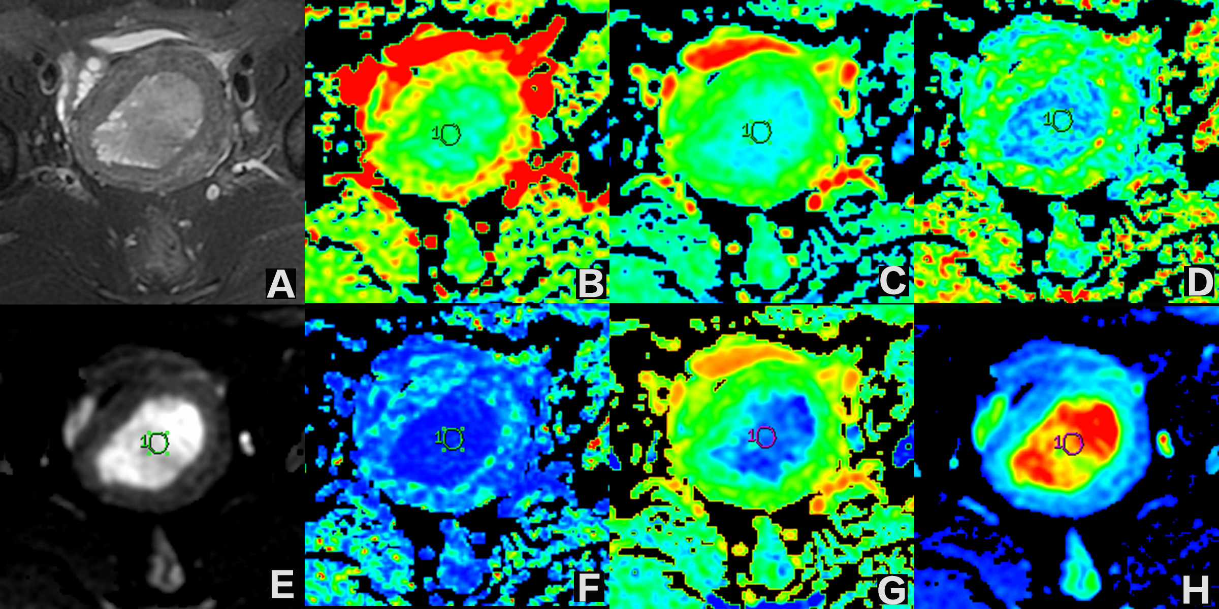
Figure 1. A-H
36 years old patients with moderately-highly differentiated endometrioid
adenocarcinoma. A.T2WI image; B. ADC diagram of DWI sequence, ADC value was
1.014×10⁻³mm2/s; C. DC avg image, DC avg value was
1.025×10⁻⁹mm2/s; D. FA image, FA value was 0.134; E. Iso
image, Iso value was 114.61; F. VRA image, VRA value was 0.020; G. Exat image,
the Exat value was 0.541; H. T2-WT image, T2-WT value was 114.615;
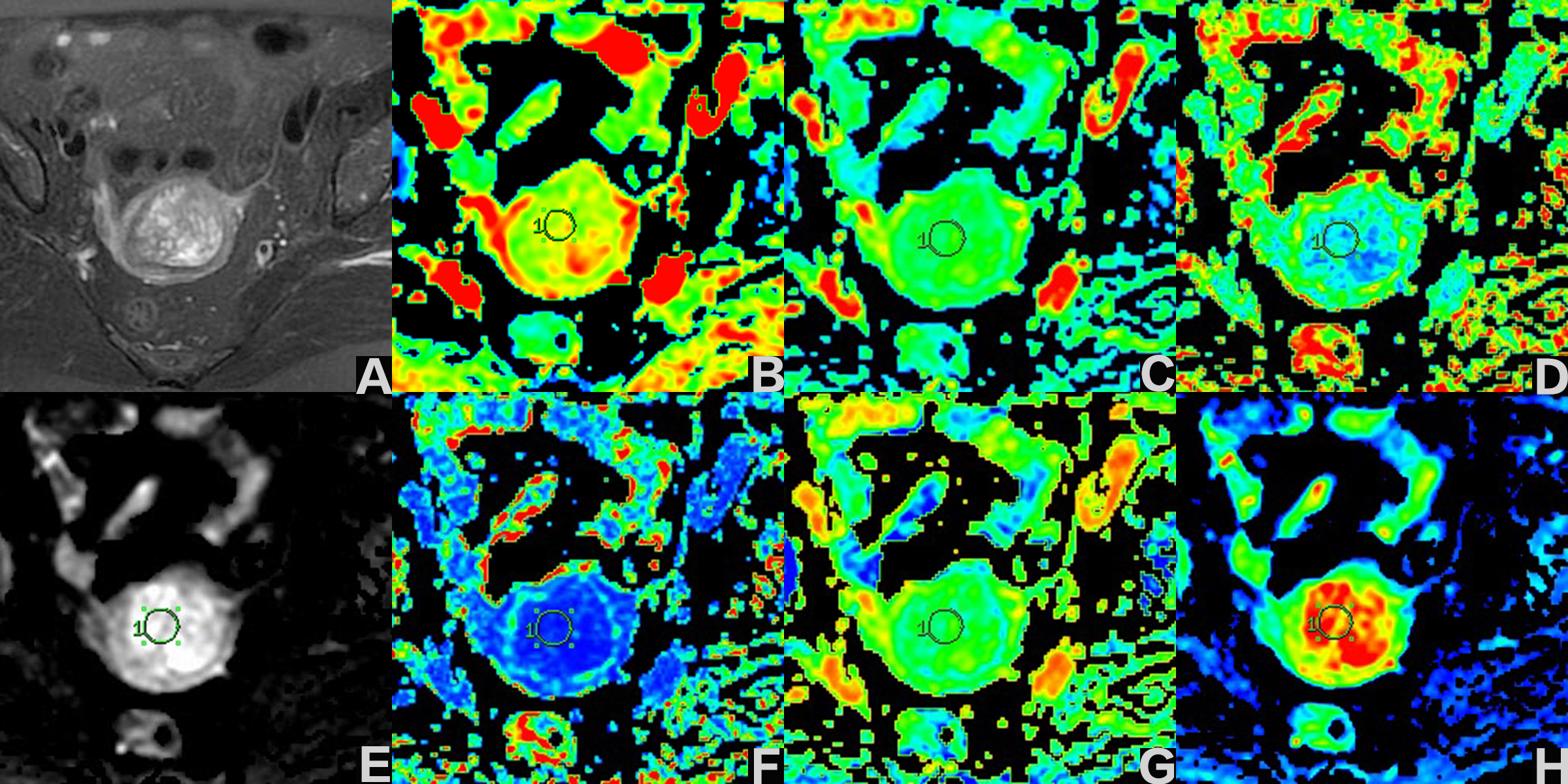
Figure
2. A-H 64 years old patients with endometrial polyp. A.T2WI image; B. ADC
diagram of DWI sequence, ADC value was 1.84×10⁻³mm2/s; C.
DC avg image, DC avg value was 1.73×10⁻⁹mm2/s; D. FA image,
FA value was 0.119; E. Iso image, Iso value was 254.755; F. VRA image, VRA
value was 0.016; G. Exat image, the Exat value was 0.355; H. T2-WT image, T2-WT
value was 254.76;