Lisan M. Morsinkhof1, Jean-Francois Witz2, Olivier Mayeur2, Anique T.M. Grob3, Frank F.J. Simonis1, and Pauline Lecomte-Grosbras2
1Magnetic Detection & Imaging, TechMed Centre, University of Twente, Enschede, Netherlands, 2Laboratoire de mécanique multiphysique multiéchelle, Université de Lille, CNRS, Centrale Lille, Lille, France, 3Multi-Modality Medical Imaging, TechMed Centre, University of Twente, Enschede, Netherlands
1Magnetic Detection & Imaging, TechMed Centre, University of Twente, Enschede, Netherlands, 2Laboratoire de mécanique multiphysique multiéchelle, Université de Lille, CNRS, Centrale Lille, Lille, France, 3Multi-Modality Medical Imaging, TechMed Centre, University of Twente, Enschede, Netherlands
Quantitative pelvic organ
mobility analysis in upright patient position is feasible. Cervix displacement is
smaller during contraction and larger during straining in supine position
compared to upright. Upright imaging may provide supplementary insight in pelvic
organ prolapse.
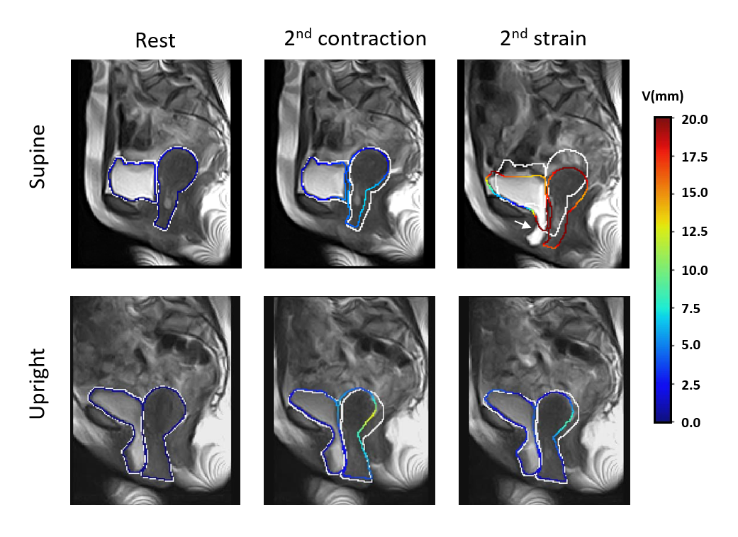
Figure 2 Vertical displacement
(V) in millimeters on the contour of pelvic organs at rest, during the second contraction
and during the second time straining. The white line represents the initial
contours, the colors represent the amount of displacement. During the second time
straining in upright position there is a mismatch between the displacement
contour and the bladder, indicated with the white arrow. This is probably
caused by in and out of plane movement of the organs during contraction and
straining, but also by the extreme mobility of the bladder in patients with
POP.
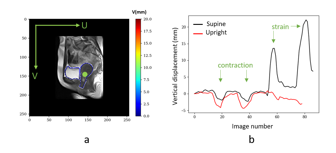
Figure 1 Displacement analysis of the cervix.
a) Point at which displacement analysis is performed (green) b) Vertical displacement
in supine (black) and upright position (red). In supine position, both
contraction and strain are clearly visible. In upright position the
displacement during contraction is larger than in supine position, and displacement
during straining is not visible. V: vertical displacement, U: horizontal
displacement