Maryam Mozaffari1,2, Nivin Nystrom1,2, Alex Li1, Miranda Bellyou1, Timothy Scholl1,2, and Robert Bartha1,2
1Robarts Research Institute, London, ON, Canada, 2Department of Medical Biophysics, Western University, London, ON, Canada
1Robarts Research Institute, London, ON, Canada, 2Department of Medical Biophysics, Western University, London, ON, Canada
The
intracellular pH of rat C6 glioma was measured at two time-points and found to
be relatively basic compared to contralateral tissue. Cariporide did not
selectively acidify this model as previously observed in mouse U87MG tumours.
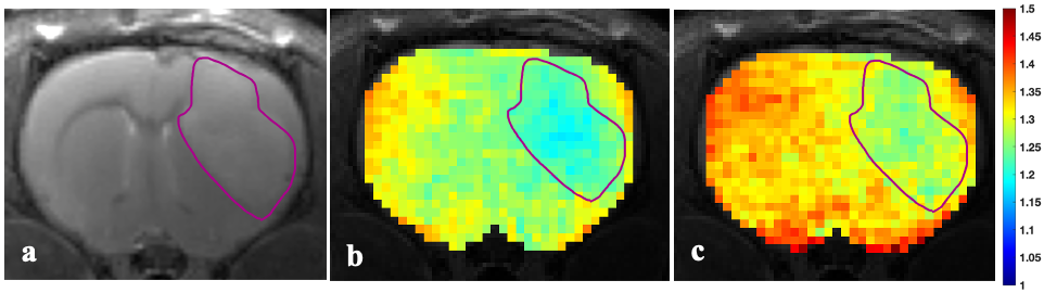
Fig. 2: a) Representative
anatomical image (T2-weighted) and superimposed colour-coded AACID
maps for b) baseline and c) 60 minutes after injection of cariporide at day 14
post-implantation of the tumour. The tumour region is highlighted by the purple
line. The average AACID value in tumour regions pre- and post-injection of
cariporide was 1.22±0.02 and 1.27±0.020, respectively.
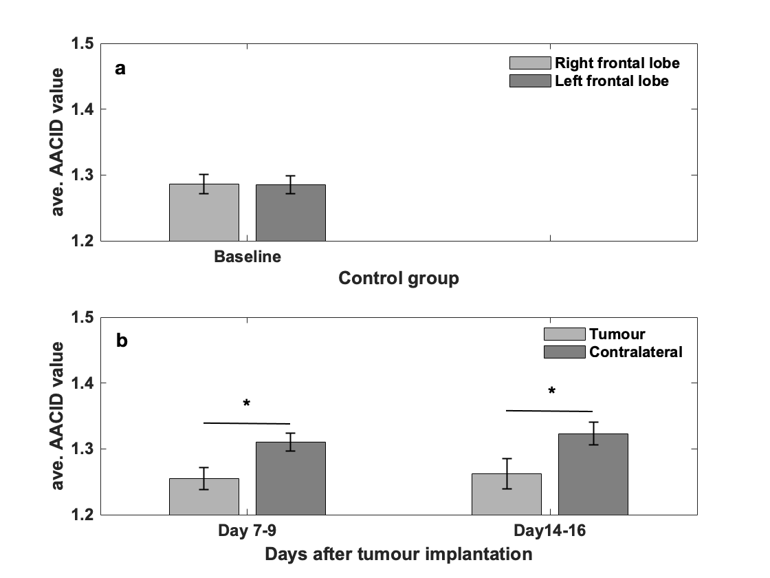
Fig. 1: The average
AACID values for a) control groups (N=15) in right and left frontal lobes. b)
experimental groups at 7-9 days (N=22) and 14-16 days (N=20) post tumour
implantation. Error bars represent the standard error of the mean. The
asterisks indicated p<0.05 in the paired t-test.