Abigail T.J. Cember1, Ravi Prakash Reddy Nanga1, Hari Hariharan1, Neil E. Wilson2, Puneet Bagga3, and Ravinder Reddy1
1Center for Magnetic Resonance and Optical Imaging, Department of Radiology, University of Pennsylvania, Philadelphia, PA, United States, 2Siemens Medical Solutions, USA, Malvern, PA, United States, 3Department of Diagnostic Imaging, St. Jude Children's Research Hospital, Memphis, TN, United States
1Center for Magnetic Resonance and Optical Imaging, Department of Radiology, University of Pennsylvania, Philadelphia, PA, United States, 2Siemens Medical Solutions, USA, Malvern, PA, United States, 3Department of Diagnostic Imaging, St. Jude Children's Research Hospital, Memphis, TN, United States
We performed volumetric (3D) gluCEST to image a slab containing the medial temporal lobe (MTL) in healthy subjects. Upon regional analysis, we find amongst other trends that the dentate gyrus has greater gluCEST contrast than neighboring regions.
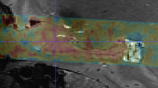
Figure 4. Example gluCEST map through medial temporal lobe slice, shown in transparent overlay on the T2-weighted structural image.
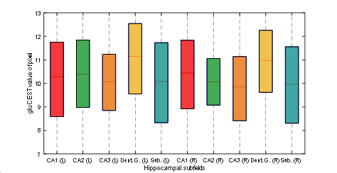
Figure 3. Comparison of gluCEST values in left and
right hippocampal subfields. This boxplot is analogous to those in Figure 1, although rather than combining the corresponding measurements on the left and right sides of the brain, we inspect each separately. It can be seen from this plot that the gluCEST distribution in the dentate gyrus is higher than in the other subfields to a corresponding degree on both sides of the brain.