Choong Heon Lee1, Piotr Walczak2, and Jiangyang Zhang1
1Radiology, NYU Medical Center, New York, NY, United States, 2University of Maryland School of Medicine, Baltimore, MD, United States
1Radiology, NYU Medical Center, New York, NY, United States, 2University of Maryland School of Medicine, Baltimore, MD, United States
ihMT can detect residual non-compact myelin in the context of cellular infiltrates in the dysmyelinating shiverer mouse model, suggesting higher sensitivity to myelin than conventional MT.
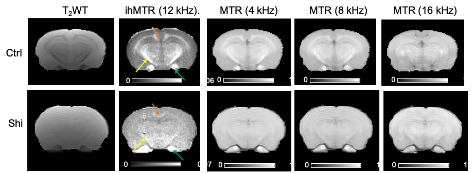
Fig. 5: Representative images of control and shiverer mouse brains. T2-weighted image (leftmost) is compared with ihMTR image at a dual offset frequency of 12 kHz as well as MTR image at offset frequencies of 4, 8, 16 kHz. Corpus callosum (orange arrow), cerebral peduncle (yellow), and trigeminal nerve (green).
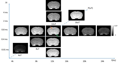
Fig. 3: Optimization of ihMTR signals in shiverer mouse brains (n=3). A series of dual offset frequencies ranging from 4k to 24k in horizontal axis; time delays from 0.01 ms to 4 ms in vertical axis; and saturation power in diagonal axis.