Yu Liu1, Junchen Li2, Ying Wang3, Naying He1, Zhijia Jin1, Pei Huang4, Shengdi Chen4, Fuhua Yan1, and Ewart Mark Haacke3
1Department of Radiology, Ruijin Hospital, Shanghai Jiao Tong University School of Medicine, Shanghai, China, 2Department of Radiology, Changshu Hospital Affiliated to Nanjing University of Chinese Medicine, Changshu, China, 3Wayne State University, Detroit, MI, United States, 4Department of Neurology, Ruijin Hospital, Shanghai Jiao Tong University School of Medicine, Shanghai, China
1Department of Radiology, Ruijin Hospital, Shanghai Jiao Tong University School of Medicine, Shanghai, China, 2Department of Radiology, Changshu Hospital Affiliated to Nanjing University of Chinese Medicine, Changshu, China, 3Wayne State University, Detroit, MI, United States, 4Department of Neurology, Ruijin Hospital, Shanghai Jiao Tong University School of Medicine, Shanghai, China
In this study, we observed that MSA with a parkinsonian variant had
significantly smaller neuromelanin (NM) volume compared with Parkinson’s
disease (PD) patients (p=0.0017) and healthy controls (HCs) (p=0.0013).
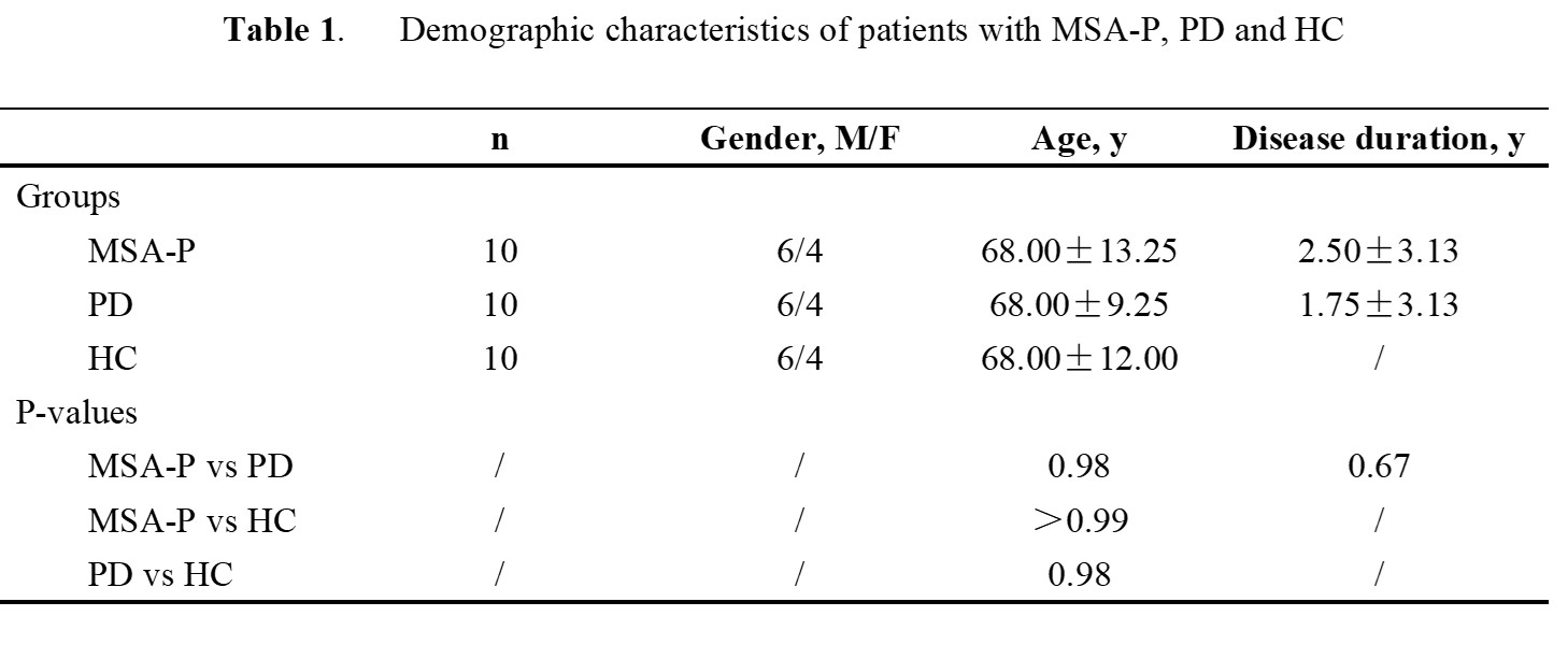
Data are presented as median ± interquartile range unless otherwise noted.
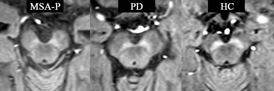
Fig. 1. MTC magnitude images
for an MSA-P patient, PD patient and HC are shown. From the MTC magnitude images of the MSA-P and PD patients, we can
easily observe the NM depigmentation with lower contrast-to-noise than HC. The
patients with MSA-P apparently suffer the worst NM degeneration.