Lihong Tang1, Hui Huang1, Miao Zhang2, Yibo Zhao3,4, Rong Guo3,4, Yudu Li3,4, Zhi-Pei Liang3,4, Wei Liu5, Yao Li1, Biao Li2, and Jie Luo1
1School of Biomedical Engineering, Shanghai Jiao Tong University, Shanghai, China, 2Department of Nuclear Medicine, Ruijin Hospital, Shanghai Jiao Tong University School of Medicine, Shanghai, China, 3Department of Electrical and Computer Engineering, University of Illinois at Urbana Champaign, Urbana, IL, United States, 4Beckman Institute for Advanced Sciences and Technology, University of Illinois at Urbana Champaign, Urbana, IL, United States, 5Department of Neurosurgery, Ruijin Hospital, Shanghai Jiao Tong University School of Medicine, Shanghai, China
1School of Biomedical Engineering, Shanghai Jiao Tong University, Shanghai, China, 2Department of Nuclear Medicine, Ruijin Hospital, Shanghai Jiao Tong University School of Medicine, Shanghai, China, 3Department of Electrical and Computer Engineering, University of Illinois at Urbana Champaign, Urbana, IL, United States, 4Beckman Institute for Advanced Sciences and Technology, University of Illinois at Urbana Champaign, Urbana, IL, United States, 5Department of Neurosurgery, Ruijin Hospital, Shanghai Jiao Tong University School of Medicine, Shanghai, China
Simultaneous high-resolution 1H-MRSI (2.0 x 3.0 x 3.0 mm3), MWF map, QSM map, and 18F-FDG-PET offer complementary information in subcortical, cortical and white matter regions for drug-resistant TLE patients, demonstrating potential for lateralization of epileptogenic zone.
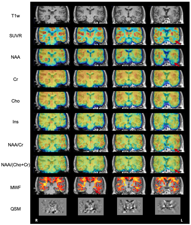
Figure 1. The coronal view high-resolution multimodal imaging of patient #3, including T1w-MPRAGE, PET-SUVR, NAA, Cr, Cho, Ins, NAA/Cr, NAA/(Cho+Cr), MWF, and QSM maps. Red arrows point to the ipsilateral temporal lobe.
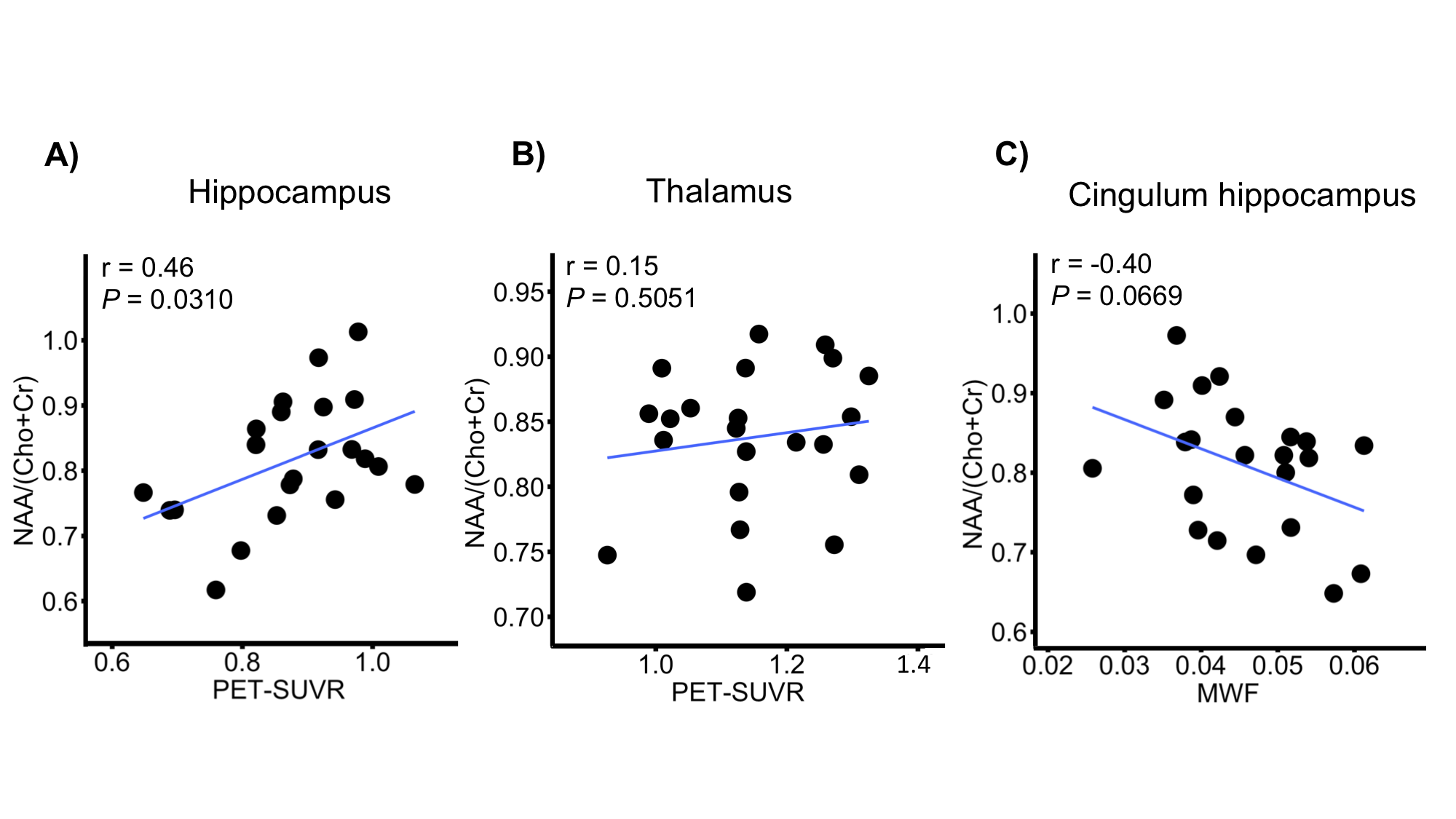
Figure 4. Relationships between A) NAA/(Cho+Cr) and PET-SUVR in hippocampus; B) NAA/(Cho+Cr) and PET-SUVR in thalamus; C) NAA/(Cho+Cr) and MWF in cingulum hippocampus. P and r values are results of Pearson’s correlation.