Jialin Hu1, Miao Zhang2, Yaoyu Zhang1, Rong Guo3,4, Yudu Li3,4, Yibo Zhao3,4, Ziyu Meng1, Biao Li2, Jun Liu5, Binyin Li5, Jie Luo1, Chao Ma6, Georges El Fakhri6, Zhi-Pei Liang3,4, and Yao Li1
1School of Biomedical Engineering, Shanghai Jiao Tong University, Shanghai, China, 2Department of Nuclear Medicine, Ruijin Hospital, Shanghai Jiao Tong University School of Medicine, Shanghai, China, 3Beckman Institute for Advanced Science and Technology, University of Illinois at Urbana-Champaign, Urbana, IL, United States, 4Department of Electrical and Computer Engineering, University of Illinois at Urbana-Champaign, Urbana, IL, United States, 5Department of Neurology and Institute of Neurology, Ruijin Hospital, Shanghai Jiao Tong University School of Medicine, Shanghai, China, 6Gordon Center for Medical Imaging, Massachusetts General Hospital, Harvard Medical School, Boston, MA, United States
1School of Biomedical Engineering, Shanghai Jiao Tong University, Shanghai, China, 2Department of Nuclear Medicine, Ruijin Hospital, Shanghai Jiao Tong University School of Medicine, Shanghai, China, 3Beckman Institute for Advanced Science and Technology, University of Illinois at Urbana-Champaign, Urbana, IL, United States, 4Department of Electrical and Computer Engineering, University of Illinois at Urbana-Champaign, Urbana, IL, United States, 5Department of Neurology and Institute of Neurology, Ruijin Hospital, Shanghai Jiao Tong University School of Medicine, Shanghai, China, 6Gordon Center for Medical Imaging, Massachusetts General Hospital, Harvard Medical School, Boston, MA, United States
We used simultaneous 3D MRSI and PET imaging to
investigate association of
neurometabolism with Aβ aggregation in
HC, MCI and AD patients. Increase in mI and decrease in NAA were found as
dementia severity increased.
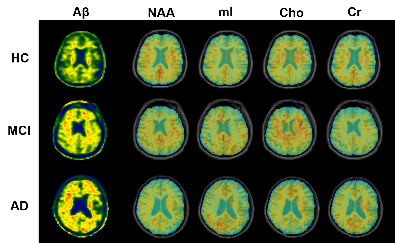
Figure 1. High-resolution metabolite maps and
PET images simultaneously acquired from a HC, an MCI patient, and an AD patient,
respectively. A global NAA reduction and mI elevation were observed with the
increased dementia severity.
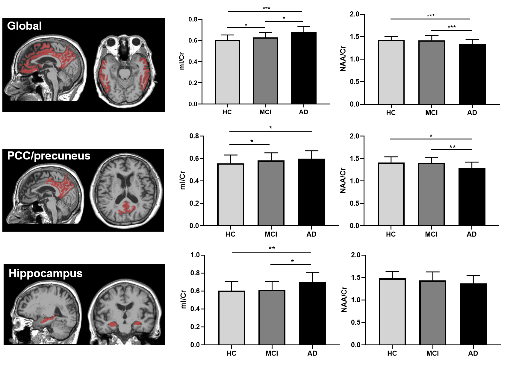
Figure 3. Comparisons of neurometabolic concentrations
among the HC, MCI, and AD groups for the global composite regions (top),
PCC/precuneus (middle) and hippocampus (bottom). * p<0.05, ** p<0.01, ***
p<0.001.