Tianyao Wang1, Danni Wang2, Yujie Hu2, Rong Guo3,4, Yudu Li3,4, Yibo Zhao3,4, Jun Liu5, Zhi-Pei Liang3,4, and Yao Li2
1Radiology Department, Shanghai Fifth People's Hospital, Fudan University, Shanghai, China, 2School of Biomedical Engineering, Shanghai Jiao Tong University, Shanghai, China, 3Department of Electrical and Computer Engineering, University of Illinois at Urbana-Champaign, Urbana, IL, United States, 4Beckman Institute for Advanced Science and Technology, University of Illinois at Urbana-Champaign, Urbana, IL, United States, 5Radiology Department, Tong Ren Hospital Shanghai Jiao Tong University School Medicine, Shanghai, China
1Radiology Department, Shanghai Fifth People's Hospital, Fudan University, Shanghai, China, 2School of Biomedical Engineering, Shanghai Jiao Tong University, Shanghai, China, 3Department of Electrical and Computer Engineering, University of Illinois at Urbana-Champaign, Urbana, IL, United States, 4Beckman Institute for Advanced Science and Technology, University of Illinois at Urbana-Champaign, Urbana, IL, United States, 5Radiology Department, Tong Ren Hospital Shanghai Jiao Tong University School Medicine, Shanghai, China
We investigated simultaneous myelin and neurometabolic alterations in acute mTBI patients. Our experimental results showed coupled myelin degradation and NAA reduction in the occipital corpus callosum.
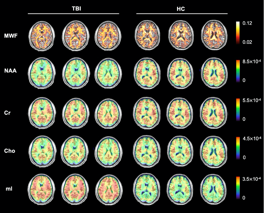
Figure 1. Reconstructed MWF and neurometabolites maps of an acute mTBI patient and a healthy control. The neurometabolites include NAA, Cr, Cho and mI. The FOV covers the whole brain (240x240x120 mm3) and the data acquisition takes 8 minutes.
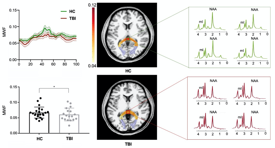
Figure 2. A reduction in MWF was observed in the occipital tract of acute mTBI patients group and the spatially resolved spectra revealed a reduction of NAA and increase of mI in the patient.