Yulu Song1, Tao Gong1, Muhammad G. Saleh2,3, Mark Mikkelsen2,3, Guangbin Wang 1, and Richard Edden2,3
1Shandong Medical Imaging Research Institute, Shandong University, jinan, China, 2Russell H. Morgan Department of Radiology and Radiological Science, The Johns Hopkins University School of Medicine, baltimore, MD, United States, 3FM Kirby Center for Functional Brain Imaging, Kennedy Krieger Institute, baltimore, MD, United States
1Shandong Medical Imaging Research Institute, Shandong University, jinan, China, 2Russell H. Morgan Department of Radiology and Radiological Science, The Johns Hopkins University School of Medicine, baltimore, MD, United States, 3FM Kirby Center for Functional Brain Imaging, Kennedy Krieger Institute, baltimore, MD, United States
We confirmed the hypothesis that a significant reduction
in the GABA+ levels in the upper brainstem regions of patients with PD compared
with the HCs.
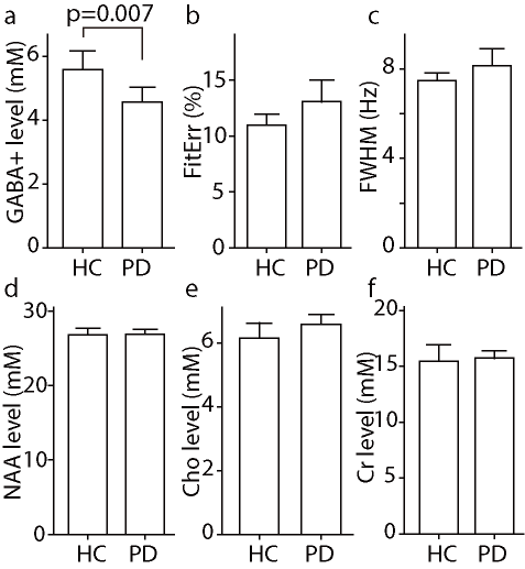
Bar Charts of
the distributions of GABA+ levels, normalized fitting errors, linewidth in Hz,
NAA, Cr, Cho levels
PD Parkinson’s disease, HC
healthy control, NAA N-acetyl aspartate, Cr creatine, Cho choline
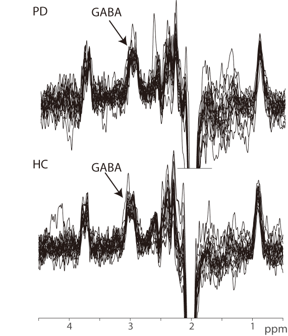
GABA+-edited
spectra in the upper brainstem of all 36 participants, showing the intended
signal at 3 ppm