William J Matloff1, Daniel J Matloff1, Arthur W Toga1, Taeuk Cheon2, Jangwook Gwak2, Yehree Kim2, Hong Ju Park2, and Hosung Kim1
1Laboratory of Neuro Imaging, USC Mark and Mary Stevens Neuroimaging and Informatics Institute, Keck School of Medicine, University of Southern California, Los Angeles, CA, United States, 2Department of Otorhinolaryngology-Head and Neck Surgery, Asan Medical Center, Seoul, Korea, Republic of
1Laboratory of Neuro Imaging, USC Mark and Mary Stevens Neuroimaging and Informatics Institute, Keck School of Medicine, University of Southern California, Los Angeles, CA, United States, 2Department of Otorhinolaryngology-Head and Neck Surgery, Asan Medical Center, Seoul, Korea, Republic of
We show the accuracy and reliability of a semi-automated approach for segmenting the modiolus, cochlear basal turn, and cochlear mid-apical turn on T2-weighted MRI images.
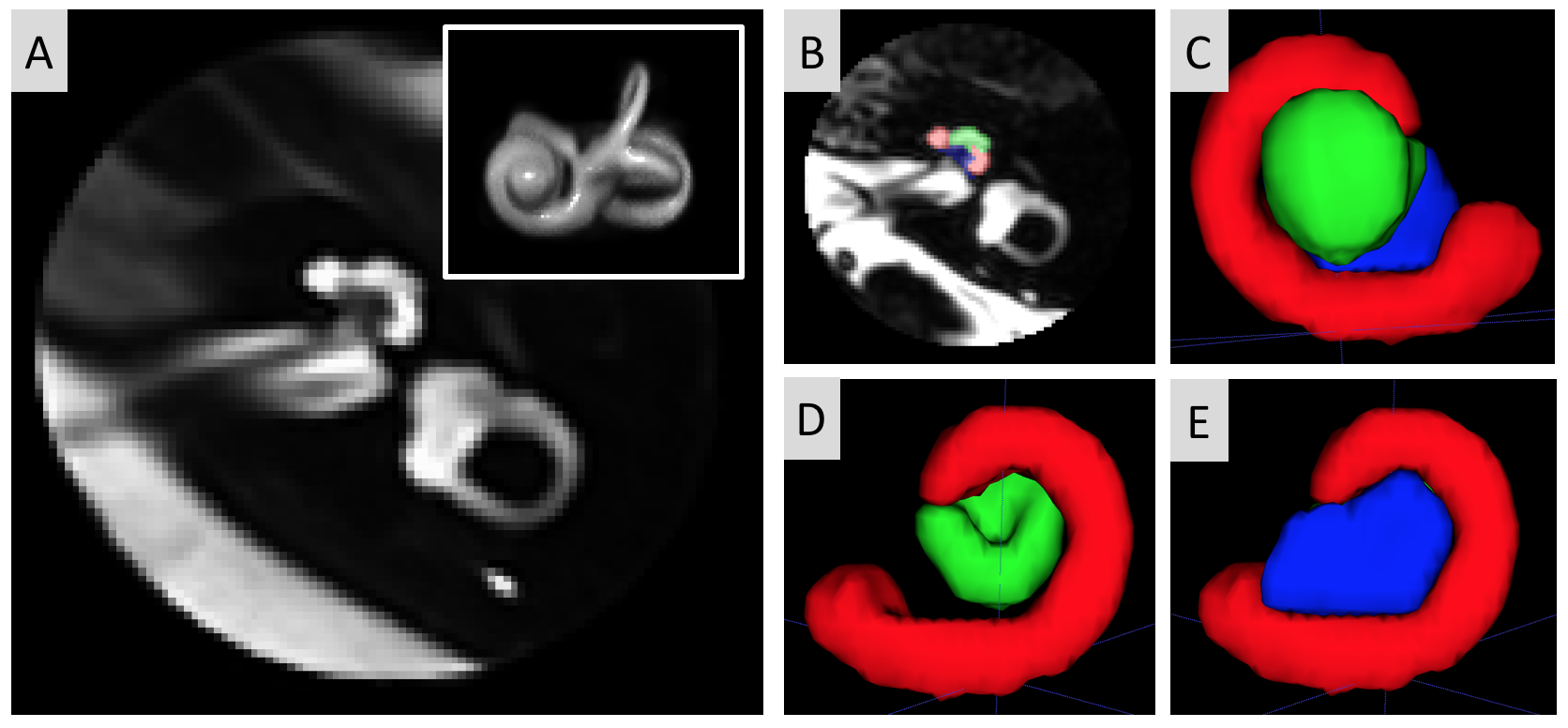
(A) Left cochlea template, with insert showing the 3D rendering of the inner ear specifically. (B) Slice of the spherical cochlea MRI region of one participant with manual segmentation overlaid. (C-E) 3D rendering of the manually segmented cochlea subregions and modiolus (red=cochlear basal turn, green=cochlear mid-apical turn, blue=modiolus) in different views.
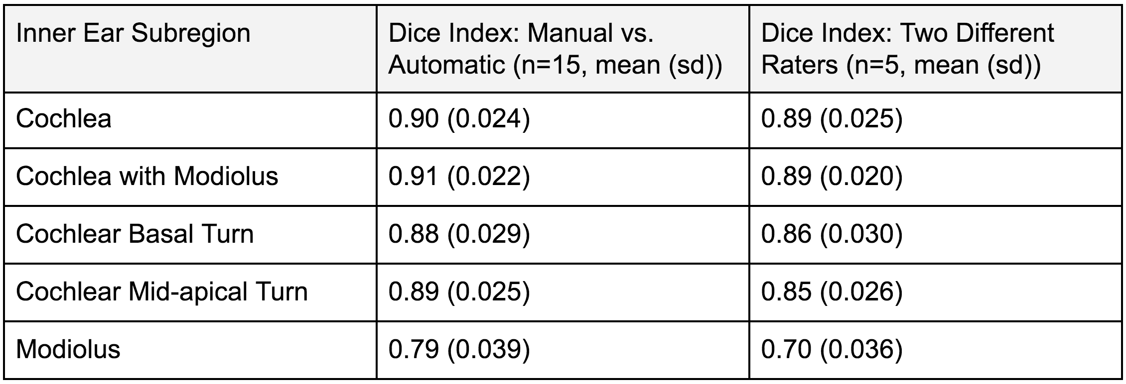
Table showing automatic segmentation agreement and inter-rater segmentation agreement.