Warda T. Syeda1, Vanessa Brait2, Alex Oman2, Charlotte Ermine 2, Jess Nithianantharajah 2, Lachlan Thompson2, Leigh A. Johnston3,4, David K. Wright5, and Amy Brodtmann2
1Melbourne Neuropsychiatry Centre, The University of Melbourne, Parkville, Australia, 2The Florey Institute of Neuroscience and Mental Health, The University of Melbourne, Parkville, Australia, 3Department of Biomedical Engineering, The University of Melbourne, Melbourne, Australia, 4Melbourne Brain Centre Imaging Unit, The University of Melbourne, Parkville, Australia, 5Department of Neuroscience, Central Clinical School, Monash University, Melbourne, Australia
1Melbourne Neuropsychiatry Centre, The University of Melbourne, Parkville, Australia, 2The Florey Institute of Neuroscience and Mental Health, The University of Melbourne, Parkville, Australia, 3Department of Biomedical Engineering, The University of Melbourne, Melbourne, Australia, 4Melbourne Brain Centre Imaging Unit, The University of Melbourne, Parkville, Australia, 5Department of Neuroscience, Central Clinical School, Monash University, Melbourne, Australia
- A multicomponent registration technique to perform non-linear registration in the presence of largely deformed ventricles in the mouse brain.
- The proposed method outperforms the single-component registration process in registering stroke data to a healthy mouse brain template.

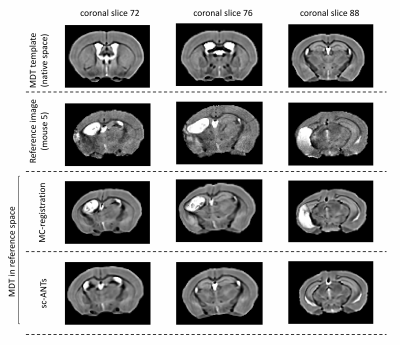
Figure 2: Exemplar coronal slices in a single mouse. Top row: MDT template (healthy template) in native space. Second row: Reference image. MDT transformed to reference image using multicomponent-ANTs (third row) and single component ANTs (last row) techniques. Multicomponent ANTs matched neuro-anatomical landmarks between MDT and individual mouse brain more robustly, with a slight overestimation of ipsilesional hippocampal boundaries.

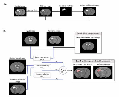
Figure 1: A) Ipsilesional ventricle contrast enhancement. A median filter is applied to stroke images. Ipsilesional ventricle intensities are upscaled 2.5x to reconstruct an enhanced image. B) MC-registration framework. Input (MDT) and reference images are affinely registered. Images are non-linearly registered using a weighted 3-component SyN transformation. Component1: cross-correlation (CC) metric (radius:r1), between MDT and reference images. Components2-3: CC metrics with radii r2 and r3, between median-filtered MDT and ventricle enhanced reference image.
