Reza Kalantar1, Susan Lalondrelle2, Jessica M Winfield1,3, Christina Messiou1,3, Dow-Mu Koh1,3, and Matthew D Blackledge1
1Division of Radiotherapy and Imaging, The Institute of Cancer Research, London, United Kingdom, 2Gynaecological Unit, The Royal Marsden Hospital, London, United Kingdom, 3Department of Radiology, The Royal Marsden Hospital, London, United Kingdom
1Division of Radiotherapy and Imaging, The Institute of Cancer Research, London, United Kingdom, 2Gynaecological Unit, The Royal Marsden Hospital, London, United Kingdom, 3Department of Radiology, The Royal Marsden Hospital, London, United Kingdom
This study introduced a novel framework to segment OARs on CT by dual-channel synthetic MRI/CT training of a multi-wavelet UNet. The results showed improved soft-tissue segmentations against a conventional UNet model.
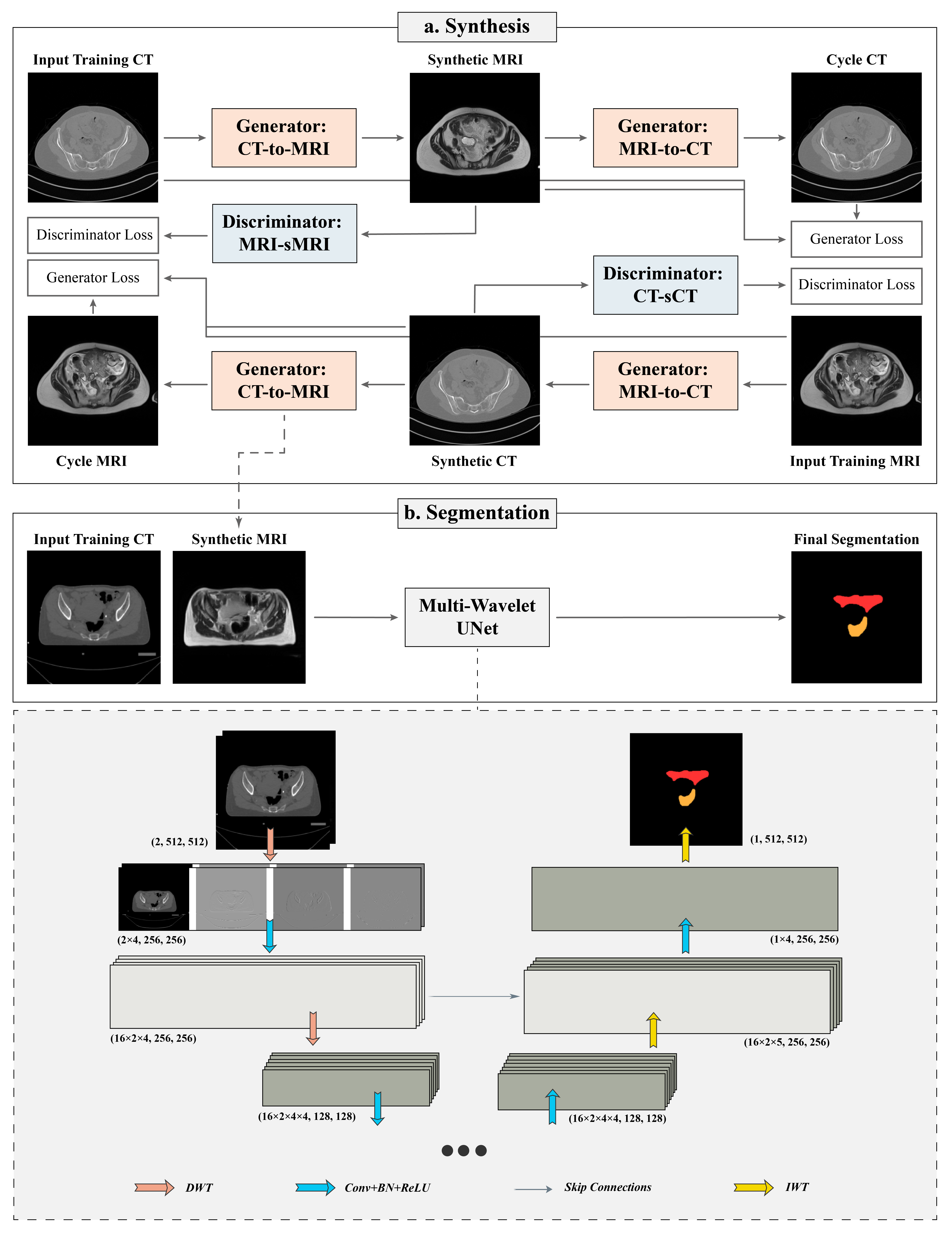
Figure 1: The proposed framework consisting of synthesis and segmentation components. (a) Synthesis: The input images in cohort A are translated to synthetic images and reconstructed back to the input domain for adversarial training. (b) Segmentation: the sMRI is generated from the input CT in cohort B to provide dual-channel input for MWUNet training. In MWUNet, DWT leads to four-fold increase in the receptive field whilst reducing the dimensionality, and IWT allows loss-less feature map reconstruction.
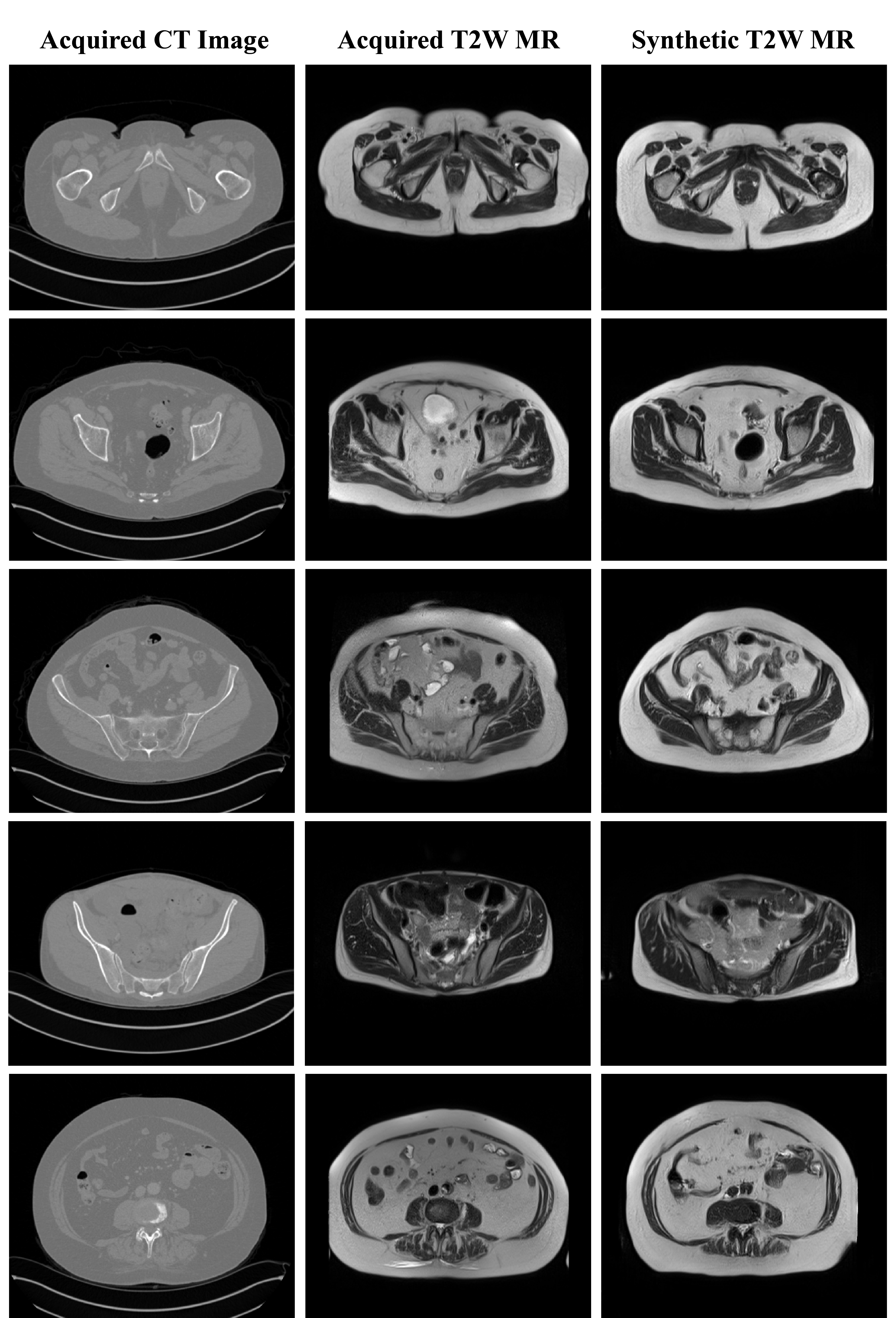
Figure 2: The synthetic T2W MRI generated from the test patients in cohort A through unpaired Cycle-GAN training. Whilst the texture and contrast were realistically predicted for structures with fixed geometries, soft-tissues with large variabilities in training images were challenging to reproduce in test sMRI. White arrow: rectum and bowel. Cyan arrow: muscle lining (fascia). Yellow arrow: examples of contrast disparity in femur and sacrum.