William Wartman1, Kyoko Fujimoto2, Mohammad Daneshzand3, Sergey Makarov1,3, and Aapo Nummenmaa3
1Electrical and Computer Engineering Department, Worcester Polytechnic Institute, Worcester, MA, United States, 2Center for Devices and Radiological Health, US Food and Drug Administration, Silver Spring, MD, United States, 3Athinoula A Martinos Center for Biomedical Imaging, Massachusetts General Hospital, Harvard Medical School, Boston, MA, United States
1Electrical and Computer Engineering Department, Worcester Polytechnic Institute, Worcester, MA, United States, 2Center for Devices and Radiological Health, US Food and Drug Administration, Silver Spring, MD, United States, 3Athinoula A Martinos Center for Biomedical Imaging, Massachusetts General Hospital, Harvard Medical School, Boston, MA, United States
For a 10-10 electrode configuration on the MIDA
model, application of electrode-specific scaling factors to the fields
calculated at the inner cortical surface for the simplified skull model can
approximate the full-model field to within 12% error.
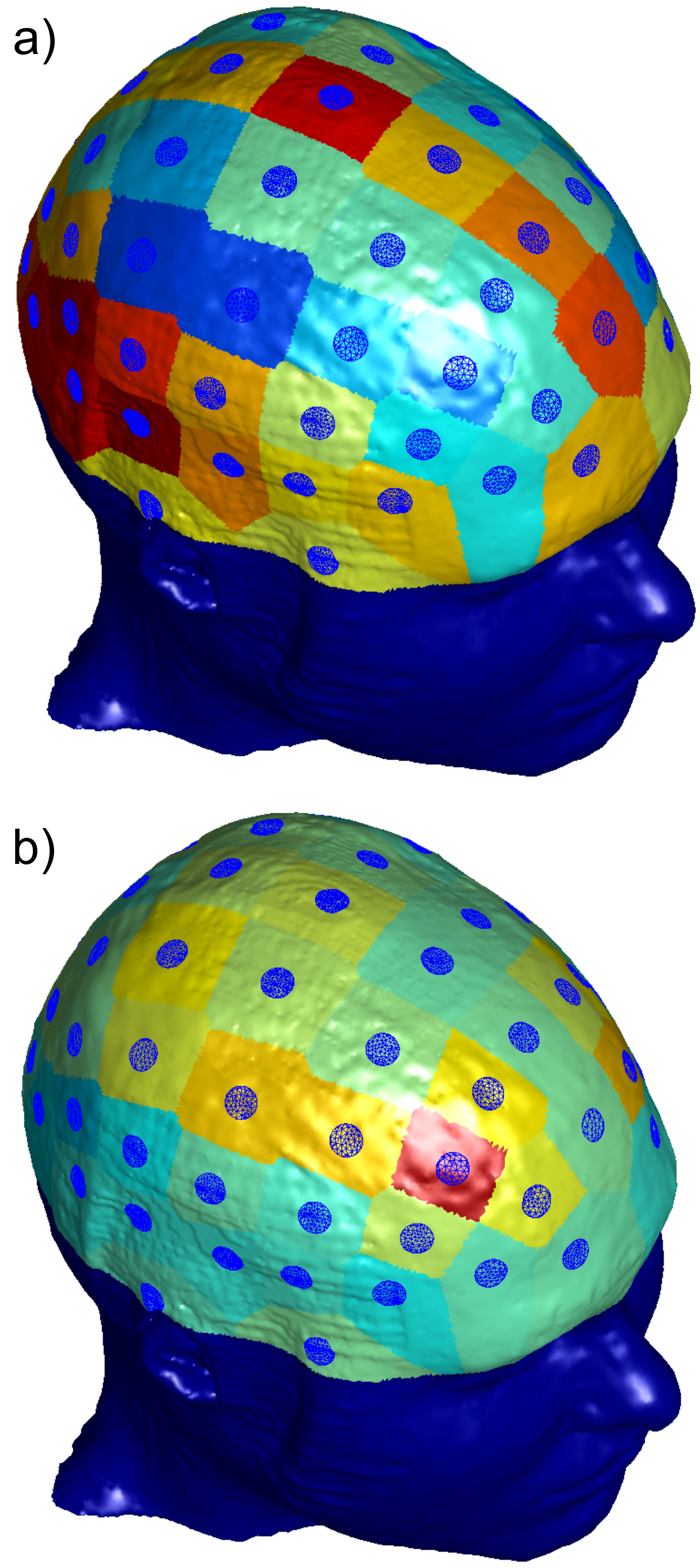
Figure 4: a): Electrode-specific corrective factors
for the total field magnitude at the WM surface, mapped to the electrode
regions on the skin surface. b): Error
between the scaled electric field magnitude for the test case and the electric
field magnitude for the base case at the white matter surface, mapped to the
electrode regions on the skin surface.
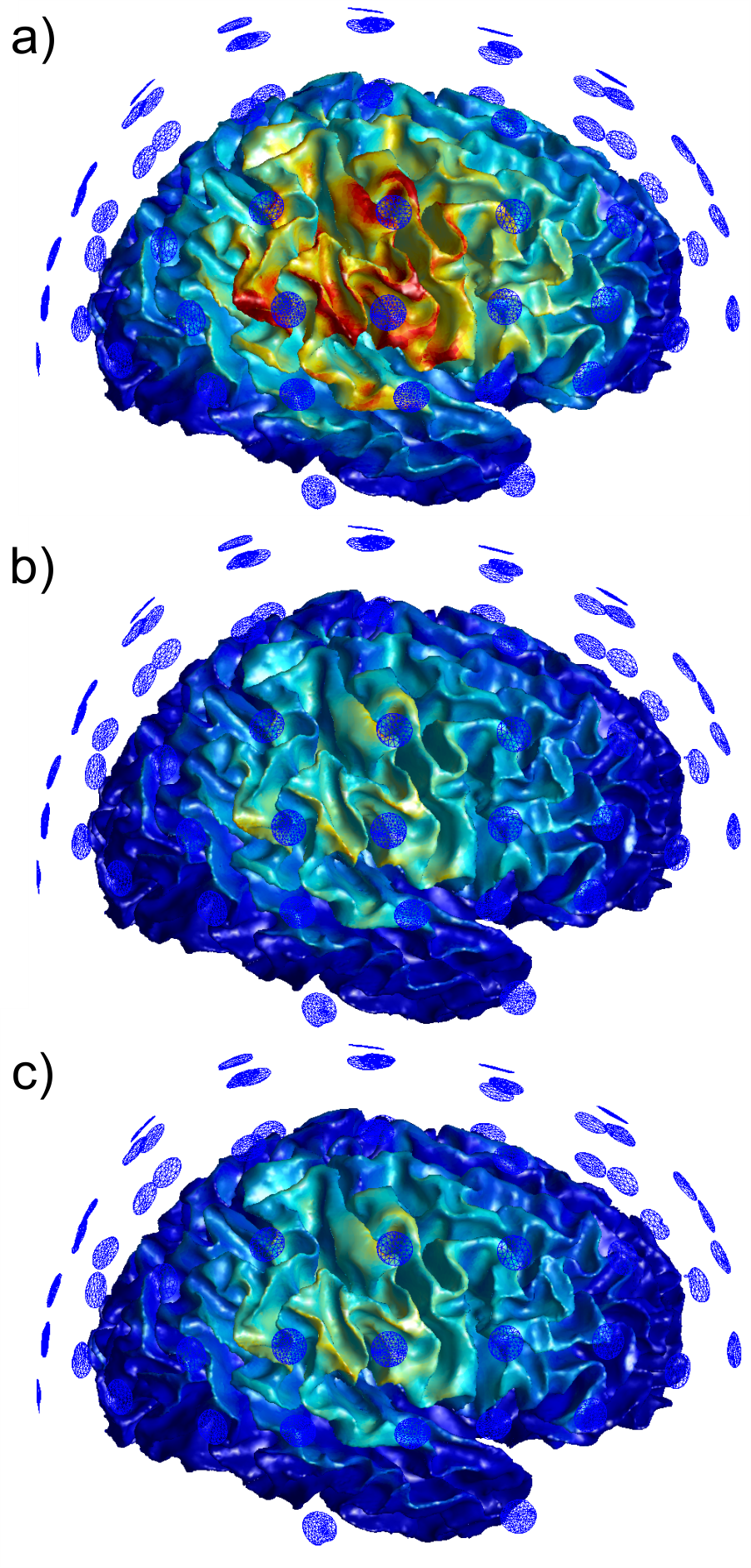
Figure 5: Magnitude of the total E-field induced by
electrode C4 just outside the WM surface.
The E-field magnitude has been clipped to 1.4 V/m in all cases. a): E-field is presented for the simplified
case in which both the diploë and dura are treated as cortical bone. b):
E-field scaled by the corrective factor is presented for the simplified
case. c): E-field is presented for the
base case where the diploë and dura are assigned their own electrical
properties.