Rafael Neto Henriques1, Marta Correia2, Maurizio Marrale3, Elizabeth Huber4, John Kruper5, Serge Koudoro6, Jason Yeatman4,7, Eleftherios Garyfallidis6, and Ariel Rokem5
1Champalimaud Research, Champalimaud Centre for the Unknown, Lisbon, Portugal, 2Cognition and Brain Sciences Unit, University of Cambridge, Cambridge, United Kingdom, 3Department of Physics and Chemistry “Emilio Segrè”, University of Palermo, Palermo, Italy, 4Institute for Learning and Brain Science and Department of Speech and Hearing, University of Washington, Seattle, WA, United States, 5Department of Psychology and eScience Institute, The University of Washington, Seattle, WA, United States, 6Department of Intelligent Systems Engineering, Luddy School of Informatics, Computing and Engineering, Indiana University Bloomington, Bloomington, IN, United States, 7Department of Pediatrics and Graduate School of Education, Stanford University, Stanford, CA, United States
1Champalimaud Research, Champalimaud Centre for the Unknown, Lisbon, Portugal, 2Cognition and Brain Sciences Unit, University of Cambridge, Cambridge, United Kingdom, 3Department of Physics and Chemistry “Emilio Segrè”, University of Palermo, Palermo, Italy, 4Institute for Learning and Brain Science and Department of Speech and Hearing, University of Washington, Seattle, WA, United States, 5Department of Psychology and eScience Institute, The University of Washington, Seattle, WA, United States, 6Department of Intelligent Systems Engineering, Luddy School of Informatics, Computing and Engineering, Indiana University Bloomington, Bloomington, IN, United States, 7Department of Pediatrics and Graduate School of Education, Stanford University, Stanford, CA, United States
DIPY provides a well-tested, well-documented, community-supported implementation of Diffusion Kurtosis Imaging (DKI), that includes a range of methods and extensions of DKI, including unique approaches to microstructure modeling and tractography.
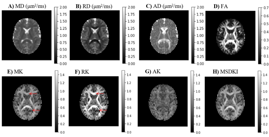
Fig. 2 - Kurtosis metrics for a representative axial slice of the CFIN data: (A) mean diffusion; (B) radial diffusivity; (C) axial diffusivity; (D) fractional anisotropy; (E) mean kurtosis; (F) radial kurtosis; (G) axial kurtosis; (H) Mean signal DKI index. The mean kurtosis tensor is relatively robust to noise-related artifacts that appear in standard MK (red arrows).
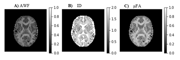
Fig.4 - Metrics from the conversion of DKI to the spherical mean technique (SMT) two compartmental model9,11: A) axonal water fraction (AWF); B) intrinsic diffusivity (ID); and C) microscopic fractional anisotropy (μFA).