Mandi Wang1, Yiang Wang1, Chia-Wei Lee2, Chien-Yuan Lin2, and Elaine Y.P. Lee1
1The University of Hong Kong, Hong Kong, Hong Kong, 2GE Healthcare, Taipei, Taiwan
1The University of Hong Kong, Hong Kong, Hong Kong, 2GE Healthcare, Taipei, Taiwan
Mean T2 value could differentiate FIGO stages in cervical
squamous cell carcinoma (SCC), and mean T2 value was higher in FIGO stage III~IV
than FIGO stage I~II.
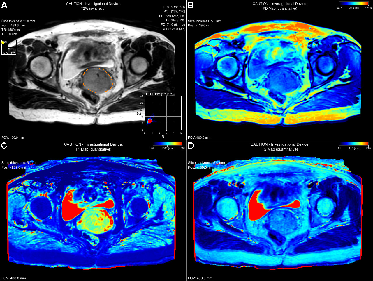
Figure 1. A 55-year-old female with squamous cell carcinoma
(SCC), FIGO stage IIIC1. Largest slice ROI was delineated on synthetic
T2-weighted image (A), the corresponding PD, T1 and T2 maps (B, C, D) were also
shown.