Xing Meng1, Ailian Liu1, Shifeng Tian1, Qinhe Zhang1, Qingwei Song1, and Jiazheng Wang2
1Department of Radiology, the First Affiliated Hospital of Dalian Medical University, Dalian, China, 2Philips Healthcare, Beijing, China
1Department of Radiology, the First Affiliated Hospital of Dalian Medical University, Dalian, China, 2Philips Healthcare, Beijing, China
The combination of APTw and IVIM
effectively enhances the differential diagnosis of EC and EP.
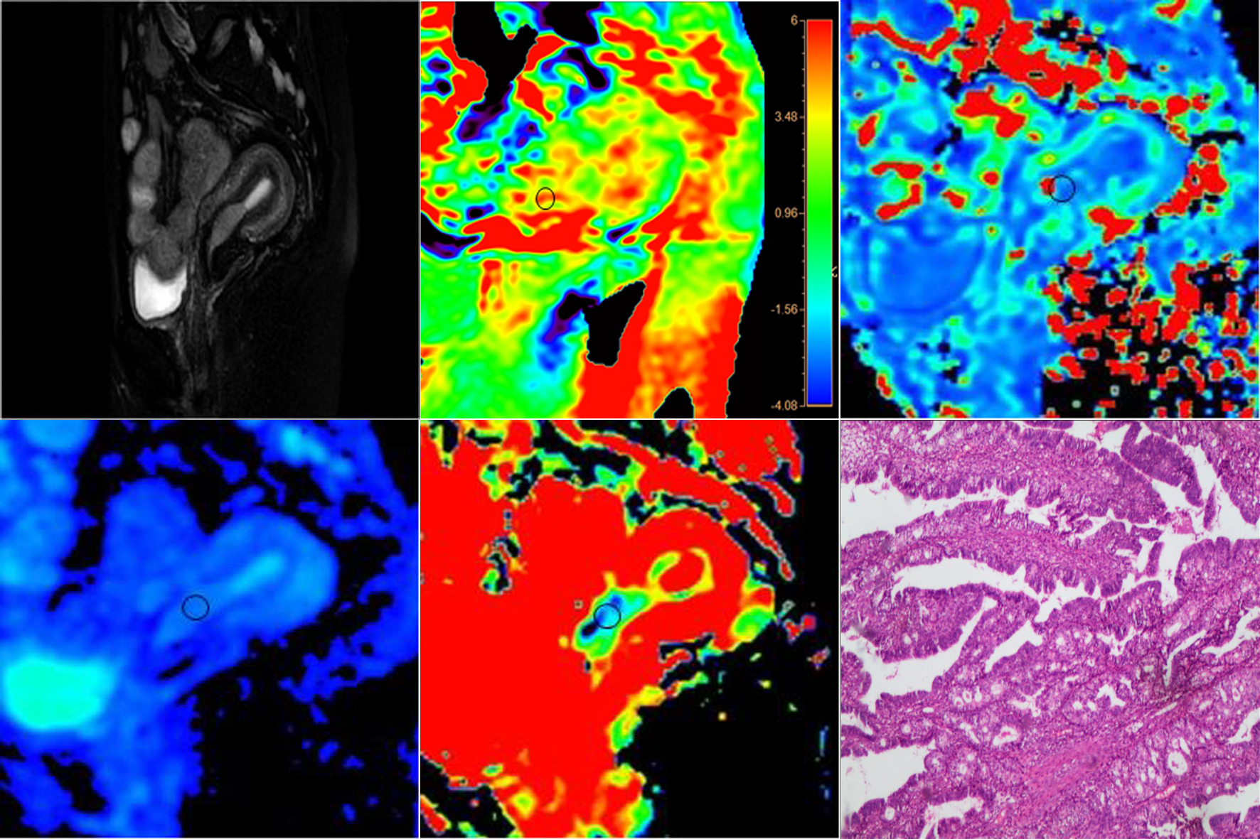
Figure
1: A 55-year-old postmenopausal patient with vaginal bleeding and endometrial
carcinoma. (A) Sag T2 SPAIR. (B) APTw and T2WI fusion map, APTw SI of 3.3%.
(C-E) IVIM post-processed pseudo-color images, the values of D*, and
the D and f values were 7.9×10⁻³mm2/s, 0.469×10⁻³mm2/s,
and 17.85%, respectively. (F) HE staining results of disease pathology
(pathological type: endometrial adenocarcinoma, magnification factor: ×40)
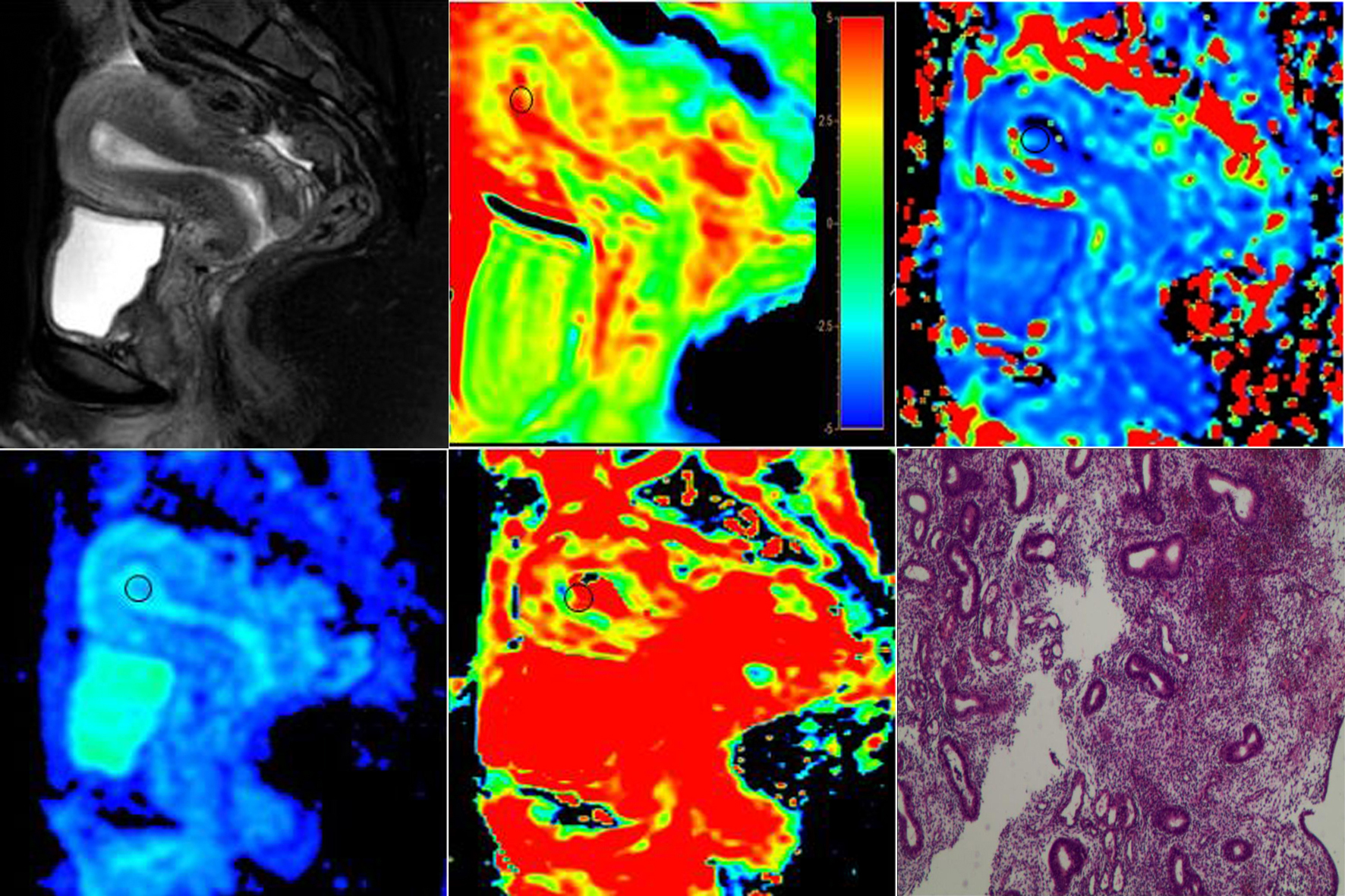
Figure
2: A 38-year-old premenopausal patient with vaginal bleeding and endometrial
polyp. (A) Sag T2 SPAIR. (B) APTw and T2WI fusion map, APTw SI of 2.35. (C-E)
IVIM post-processed pseudo-color images, the values of D*, D, and f
were 7.0×10⁻³mm2 /s, 0.988×10⁻³mm2/s,
and 51.8%, respectively. (F) HE staining results of disease pathology
(magnification factor: ×40)