Nan Meng1, Zhun Huang2, Pengyang Feng2, Ting Fang1, Fangfang Fu1, Yaping Wu1, Wei Wei1, Xuejia Wang3, Kaiyu Wang4, and Meiyun Wang*1
1Department of Radiology, Zhengzhou University People’s Hospital & Henan Provincial People’s Hospital, Zhengzhou, China, 2Department of Radiology, Henan University People’s Hospital & Henan Provincial People’s Hospital, Zhengzhou, China, 3Department of MRI, The First Affiliated Hospital of Xinxiang Medical University,, Weihui, China, 4GE Healthcare, MR Research China, BeiJing, China
1Department of Radiology, Zhengzhou University People’s Hospital & Henan Provincial People’s Hospital, Zhengzhou, China, 2Department of Radiology, Henan University People’s Hospital & Henan Provincial People’s Hospital, Zhengzhou, China, 3Department of MRI, The First Affiliated Hospital of Xinxiang Medical University,, Weihui, China, 4GE Healthcare, MR Research China, BeiJing, China
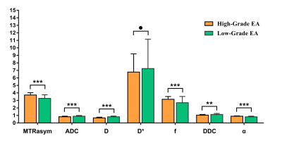
Both multimodel DWI and APTWI can be used to estimate the histological grade and Ki-67 index of EA, and the combination of a high MTRasym(3.5 ppm) and low D may be an effective imaging marker for predicting the grade of EA.

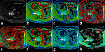
Figure.1. Low-grade EA (grade 1, FIGO IB, Ki-67 = 10%) in a 49-year-old woman (arrowheads). (a) Map of DWI (b = 1000s/mm2). (b) Pseudo colored map of ADC, (c) Pseudo colored map of D, (d) Pseudo colored map of D*, (e) Pseudo colored map of f, (f) Pseudo colored map of DDC, (g) Pseudo colored map of α, (h) Pseudo colored map of MTRasym (3.5ppm).

